
Loading...
![[Translate to chinese:] Video Talk by Kurt Thorn: The Abbe Diffraction Experiment](/fileadmin/_processed_/2/b/csm_Video_Talk_by_Kurt_Thorn_1301b7b8d9.png "[Translate to chinese:] Video Talk by Kurt Thorn: The Abbe Diffraction Experiment")
Kurt Thorn的视频讲座:阿贝衍射实验
本讲座介绍了恩斯特-阿贝的著名实验,该实验显示了试样对光线的衍射(以及与照明光线的干涉)是如何产生图像的,以及衍射光线的收集是如何定义显微镜的分辨率的。这些概念是通过使用衍射光栅作为样本,以及可视化和比较后焦平面的衍射图案以及图像平面的图像来展示的。
Loading...
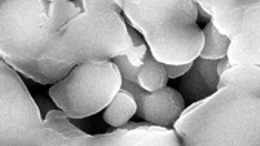
Porous Ceramics - Sample Preparation for SEM
Application Note for Leica EM RES102 - Ceramic membrane filters with pore sizes down to a few nanometres must be investigated in cross-section with regard to the structure of the pores. The smallest…
Loading...

具有可实现更高效检查和质量控制的通用照明和各种对比方法的数字显微镜
利用能够实现多种对比度方法的最先进数字显微镜,例如Leica DVM6,对于检查、质量控制和故障分析非常有效。这些对比度方法可以更容易、更快速地检测出产品或组件表面的瑕疵或缺陷。
Loading...

Chronic Inflammation Under the Microscope
In the course of chronic inflammation certain body areas are recurrently inflamed. This goes along with many human diseases. With the help of widefield light microscopy, the underlying processes can…
Loading...
什么是OCT?
光学相干断层扫描(OCT)是一种无创、非接触式成像方式,可用于显示并监测生物组织的形态变化。OCT利用低相干干涉原理,可生成横断面视图,以揭示感兴趣组织的亚表面细节。在大部分常见眼科应用中,OCT系统通过近红外光生成角膜、虹膜、晶状体、玻璃体和视网膜等组织微结构的高分辨率体积图像。这些图像可加强对病理状况的理解,例如青光眼、老年性黄斑变性或糖尿病视网膜病变。
Loading...
![[Translate to chinese:] Wifi education solutions](/fileadmin/_processed_/c/e/csm_csm_wifi-education-solutions2_03_0a47178324_a83e70edc8.jpg "[Translate to chinese:] Wifi education solutions")
选择学生显微镜需要考虑的因素
对于教师来说,选择教育显微镜并非易事。显微镜必须经得起并非总是小心翼翼的双手的日常使用,必须能够持续运行,还必须符合预算要求。尤其是学生用显微镜,实用性方面起着重要作用: 尺寸、重量、布线和设计在日常使用中非常重要,甚至在决定使用显微镜的设备和附件之前就应考虑到这一点。如果选择得当,教育显微镜将为大中小学的年轻人打开一扇通往微小细节的宇宙之窗,让他们对科学产生足够的兴趣,并将其作为自己的职业。
Loading...

Gene Editing with CRISPR/Cas9 - Breakthrough in Genome Engineering
The CRISPR/Cas9 system is one of several different bacterial systems for defense against viral attacks. It consists of two main components. One is a small piece of RNA which binds to the viral target…
Loading...
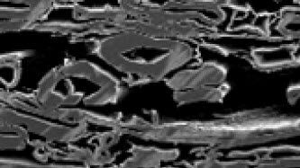
Paper Samples - Sample Preparation for SEM
Application Note for Leica EM RES102 - A coated paper sample has been prepared with ion beam slope cutting in order to test the procedure with regard to its applicability. With the use of ion beam…
Loading...

Imaging and Analyzing Zebrafish, Medaka, and Xenopus
Discover how to image and analyze zebrafish, medaka, and Xenopus frog model organisms efficiently with a microscope for developmental biology applications from this article.