
Loading...
 or Minor’s syndrome")
Minor’s Syndrome Surgical Intervention by Prof. Vincent Darrouzet
Minor’s disease, also called Superior Semicircular Canal Dehiscence (SSCD) or Minor’s syndrome, is a rare disorder of the inner ear that affects hearing and balance. The disease is characterized by…
Loading...
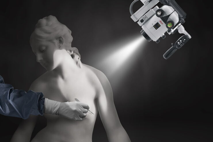
如何为重建手术选择显微镜
整形和重建手术需要良好的可视化来修复复杂和精细的结构。肿瘤重建手术是精细的手术之一,包括乳房重建、淋巴重建和头颈重建(颌面外科)。从皮肤移植到游离皮瓣,这些手术需要使用多种技术。手术显微镜可以帮助解决手术过程中的一些困难。发现五个决定性的特点来指导您选择整形和重建手术显微镜。
Loading...

Advances in Oncological Reconstructive Surgery
Decision making and patient care in oncological reconstructive surgery have considerably evolved in recent years. New surgical assistance technologies are helping surgeons push the boundaries of what…
Loading...
![[Translate to chinese:] H&E stained specimen, 20x magnification](/fileadmin/_processed_/1/6/csm_H_E_stained_specimen_20x_10_K3_121c9519a6.jpg "[Translate to chinese:] H&E stained specimen, 20x magnification")
诊断时间在临床病理学检查中至关重要
病理学家使用显微镜检查样本以发现组织和体液中存在的异常。他们的检查发现或推论对治疗决策有非常大的影响。Penelope Zorzi博士拥有免疫学、病理学和基因组学专业背景,是徕卡显微系统健康与临床显微镜术产品工作流经理。她了解病理学家的工作职责并介绍了医生、科学家和病理学实验室化验员的工作。
Loading...
A Versatile Palette of Fluorescent Probes
Researchers at the Max Planck Institute for Medical Research in Heidelberg have developed a general strategy to synthesize live-cell compatible fluorogenic probes, and the result are the new MaP (Max…
Loading...
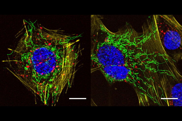
![[Translate to chinese:] Dual color volume rendering of Drp1 oligomers (green) and mito OM (red) in a live U2OS cell](/fileadmin/_processed_/1/2/csm_Dual_color_volume_rendering_in_a_live_U2OS_cell_64e6b2186b.jpg "[Translate to chinese:] Dual color volume rendering of Drp1 oligomers (green) and mito OM (red) in a live U2OS cell")
多色四维超分辨光片显微镜
人工智能显微术研讨会主要关注和讨论显微术和生物医学成像领域的最新人工智能技术和工具。在该科学演示中,Yuxuan Zhao展示了如何通过渐进式深度学习策略并结合“双环调制的SPIM”设计改善活细胞中的细胞器三维成像。
Loading...
![[Translate to chinese:] Colon adenocarcinoma with 13 biomarkers shown](/fileadmin/_processed_/4/9/csm_Colon_adenocarcinoma_with_13_biomarkers_shown_42edcfed0a.jpg "[Translate to chinese:] Colon adenocarcinoma with 13 biomarkers shown")
利用Cell DIVE 在单细胞水平上进行超复杂癌症组织分析
能够研究淋巴瘤细胞的异质性如何受到细胞对其微环境反应的影响,尤其是在突变、转录组和蛋白质水平上。蛋白质表达研究提供了有关细胞相互作用性质和蛋白质表达水平的最相关信息。超复合工作流程可用于研究同一癌症组织中的多种蛋白质。
Loading...

精确分析宽视野荧光图像
利用荧光显微镜的特异性,即便是使用厚样品和大尺寸样品,研究人员也能够快速轻松地准确观察和分析生物学过程和结构。然而,离焦荧光会提高背景荧光,降低对比度,影响图像的精确分割。THUNDER 与Aivia 的组合可以有效解决这一问题。前者可以消除图像模糊,后者会使用人工智能技术自动分析宽视野图像,提高操作速度和精确性。下面,我们来详细了解下这一协作方法。
Loading...

光学相干断层扫描(OCT)引导下视网膜手术的临床研讨会
在本记录中,来自新加坡某眼科中心的A教授和来自西班牙巴塞罗那某儿童医院的B医生分享了他们使用眼科显微镜所提供的术中OCT行视网膜手术的技术经验。他们报告了从常规黄斑裂孔手术到基因治疗的多个感兴趣儿科病例。