
Loading...

斑马鱼大脑高分辨率全器官成像
结构信息是理解复杂生物系统的关键,而脊椎动物的中枢神经系统是最复杂的生物结构之一。要想从发育中的斑马鱼身上分离出一个完整的大脑,我们需要覆盖大约10平方毫米的区域,深度在毫米范围内。通常,低倍透镜不能提供足够的分辨率来揭示神经组织中复杂结构之间的相互作用。此外,由于散射过程,使用共聚焦显微镜在致密生物组织内成像深度通常限制在大约10微米。
Loading...
Improving RNA Analysis with Laser Microdissection
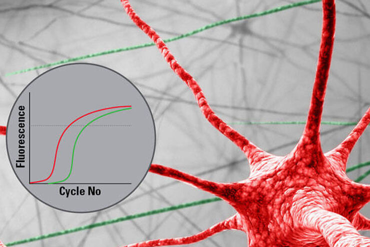
Parkinson’s disease is a progressive neurodegenerative disorder connected with cell death of dopamine-releasing neurons in the brain. Differences in gene expression between individual…
Loading...
How to improve your Alzheimer Protein Analysis with Laser Microdissection
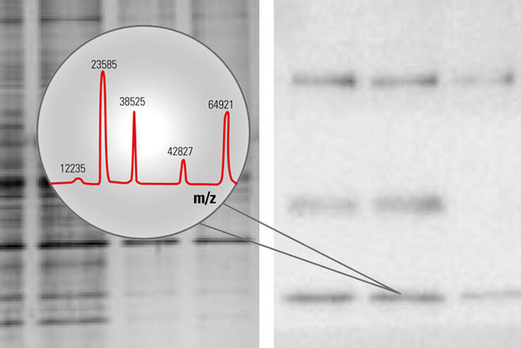
Brain Research: Collect pure starting material for proteomics - Improve your workflow with Laser Microdissection - Many brain diseases result from protein malfunction, misfolding and agglutination.…
Loading...

Studying Caenorhabditis elegans (C. elegans)
Find out how you can image and study C. elegans roundworm model organisms efficiently with a microscope for developmental biology applications from this article.
Loading...
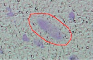
The Morbus Parkinson Puzzle
A characteristic sign of M. Parkinson is the deterioration of dopaminergic neurons in the mid-brain, specifically in the substantia nigra (SN, black substance). Different causes and forms of this…
Loading...

Ratiometric Imaging
Many fundamental functions of a cell strongly depend on delicate, but nevertheless dynamic balances of ions (e.g. calcium, magnesium), voltage potentials and pH between the cell’s cytosol and the…
Loading...
New Standard in Electrophysiology and Deep Tissue Imaging
The function of nerve and muscle cells relies on ionic currents flowing through ion channels. These ion channels play a major role in cell physiology. One way to investigate ion channels is to use…