
![[Translate to chinese:] Spheroid stained with Cyan: Dapi nuclear countertain; Green AF488 Involucrin; Orange AF55 Phalloidin Actin; Magenta AF647 CK14.](/fileadmin/_processed_/6/8/csm_Spheroid_stained_with_Cyan_4color_overlay_76cb6e4d04.jpg "[Translate to chinese:] Spheroid stained with Cyan: Dapi nuclear countertain; Green AF488 Involucrin; Orange AF55 Phalloidin Actin; Magenta AF647 CK14.")
Loading...

Virtual Reality Showcase for STELLARIS Confocal Microscopy Platform
In this webinar, you will discover how to perform 10-color acquisition using a confocal microscope. The challenges of imaged-based approaches to identify skin immune cells. A new pipeline to assess…
Loading...
")
Wt1 Genes Can Induce a Cardiomyocyte to Epicardial-like Cell Fate Transition
From this study, it was concluded that Wt1 plays a yet undescribed role for cardiomyocyte differentiation by repressing chromatin opening at specific genomic loci and that sustained ectopic expression…
Loading...
![[Translate to chinese:] Dual color volume rendering of Drp1 oligomers (green) and mito OM (red) in a live U2OS cell](/fileadmin/_processed_/1/2/csm_Dual_color_volume_rendering_in_a_live_U2OS_cell_64e6b2186b.jpg "[Translate to chinese:] Dual color volume rendering of Drp1 oligomers (green) and mito OM (red) in a live U2OS cell")
多色四维超分辨光片显微镜
人工智能显微术研讨会主要关注和讨论显微术和生物医学成像领域的最新人工智能技术和工具。在该科学演示中,Yuxuan Zhao展示了如何通过渐进式深度学习策略并结合“双环调制的SPIM”设计改善活细胞中的细胞器三维成像。
Loading...

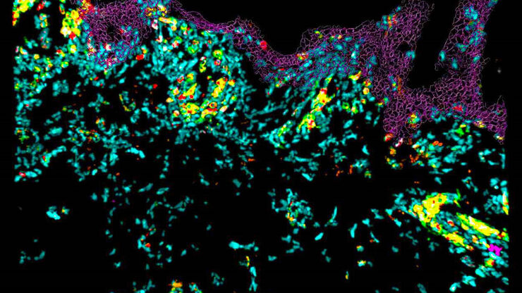
Imaging of Anti-Cancer Drug Uptake in Spheroids using DLS
Spheroid 3D cell culture models mimic the physiology and functions of living tissues making them a useful tool to study tumor morphology and screen anti-cancer drugs. The drug AZD2014 is a recognized…
Loading...
透明介质对组织透明度和收缩的影响
本研究通过比较新鲜解剖的双翅目昆虫脑与其透明化处理后的等效物,全面评估了不同透明化介质对组织透明度和收缩率的影响。组织透明化处理结合光片显微技术,已成为全器官三维成像和定量的强大工具。由于组织透明化处理有助于在亚细胞水平上对完整组织进行光学成像,它有潜力揭示如大脑等复杂器官以前未见的细节。为了对厚样本进行高分辨率成像,透明化介质需要与高数值孔径(NA)物镜使用的浸没液具有匹配的高折射率。这一点对于…
Loading...
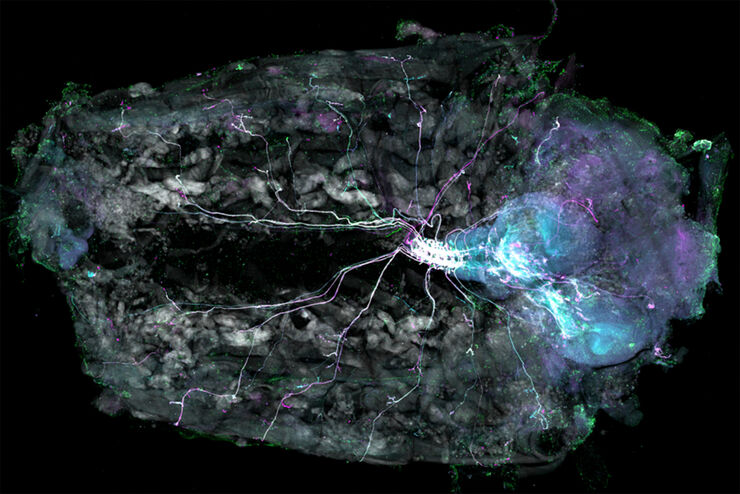
Understanding Motor Sequence Generation Across Spatiotemporal Scales
We have developed a microscopy-based pipeline to characterize a developmentally critical behavior at the pupal stage of development, called the ecdysis sequence. We study brain-wide neuronal activity…
Loading...

Confocal and Light Sheet Imaging with STELLARIS DLS
Optical imaging instrumentation can magnify tiny objects, zoom in on distant stars and reveal details that are invisible to the naked eye. But it notoriously suffers from an annoying problem: the…