Webinars

最新市场活动资讯

全部活动

Developing embryos of different species at different stages during the elongation of their posterior body axis, from left to right in developmental time. The labelled regions in red depict a region of undifferentiated cells called the tailbud, with the corresponding region generated from that tissue shaded in grey. Upper row: lamprey; middle row: catshark; bottom row, zebrafish. This figure has been adapted from the following publication: Steventon, B., Duarte, F., Lagadec, R., Mazan, S., Nicolas, J.-F., & Hirsinger, E. (2016). Species tailoured contribution of volumetric growth and tissue convergence to posterior body elongation in vertebrates. Development, 2016. 143(10):1732-41

如何研究胚胎发育过程中的基因调控网络

请与 Andrea Boni 博士一起参加本次点播网络研讨会,探索光片显微镜如何彻底改变发育生物学。这种先进的成像技术可对三维样本进行高速、容积式实时成像,且光毒性极低。通过用户实例了解光片显微镜如何增强我们对肠道和大脑类器官发育的理解,并深入了解徕卡显微系统公司的 Viventis Deep 显微镜背后的技术及其在长期成像中的应用。
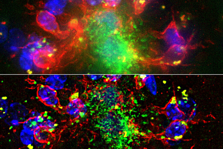
The role of extracellular signalling mechanisms in the correct development of the human brain

在神经发育过程中,细胞是如何相互交流的?

细胞间通信是大脑发育过程中一个必不可少的过程,它受到多种因素的影响,包括细胞的形态、粘附分子、局部细胞外基质和分泌囊泡。在本次网络研讨会上,您将了解到对这些机制更深入的理解是如何推动对神经发育障碍的理解的。
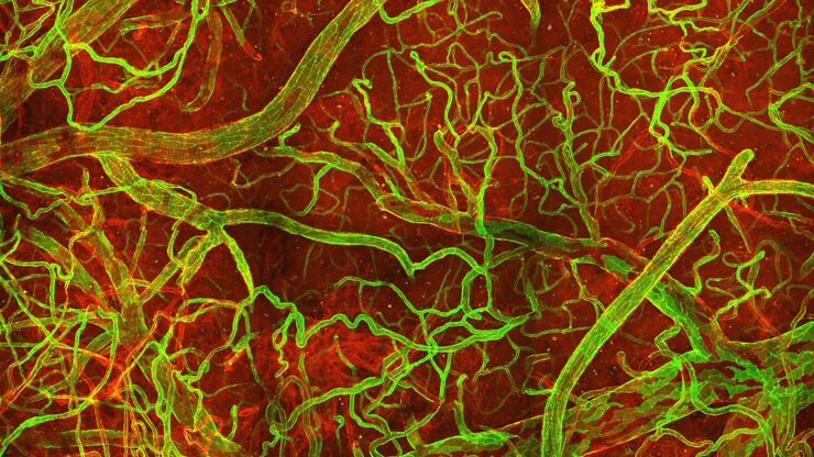
In vivo imaging of a mouse pial and cortical vasculature through a glass window (ROSAmT/mG::Pdgfb-CreERT2 mouse meningeal and cortical visualization following tamoxifen induction and craniotomy). Courtesy: Thomas Mathivet, PhD

神经血管病理学之窗

探索先天性免疫如何在神经血管病变后持续产生有害影响,以及能够对这些事件进行纵向研究的技术发展。
THY1-EGFP labeled neurons in mouse brain processed using the PEGASOS 2 tissue clearing method, imaged on a Leica confocal microscope. Neurons were traced using Aivia’s 3D Neuron Analysis – FL recipe. Image credit: Hu Zhao, Chinese Institute for Brain Research.

借助人工智能,揭示复杂而密集的神经元图像中的洞察

神经元的3D形态学分析通常需要使用不同的成像模式,捕捉多种类型的神经元,并在各种密度下相连的传统Leica SP8显微镜采集多达解神经元的形态,这对许多研究人员来说仍然是一个耗时的挑战。

Computational Clearing - 增强3D标本成像

本次网络研讨会旨在阐明有助于THUNDER显微成像仪实现三维样品可视化的关键规格,并改进研究人员的成像相关工作流程。
Scroll to top