
生命科学研究

生命科学研究

生命科学研究

在生命科学研究中心,您可以掌握最新的关于先进显微镜、成像技术、电镜样品制备和图像分析的前沿应用和创新,涵盖的主题包括细胞生物学、神经科学和癌症研究。希望在这里可以帮助您提升研究能力和精进显微镜在各个科学领域实际应用,并了解徕卡如何通过精确的可视化、图像解读和推进研究进展来赋能您的工作。
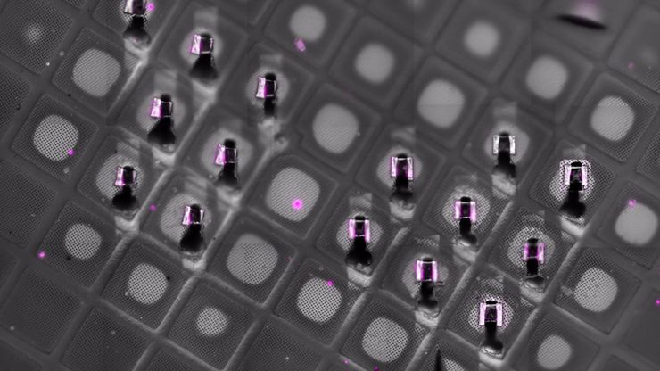
Cryo-confocal fluorescence imaging of the milled lamella on the TEM grid after cryo-FIB preparation. Residual fluorescence signal confirms that the targeted region of interest—containing malaria parasite structures—has been successfully retained during lamella preparation, supporting accurate correlation between cryo-fluorescence microscopy and cryo-electron microscopy workflows.

Fluorescence-Guided Cryo-ET of Malaria Parasites

Correlative cryo workflow with cryo-planing and cryo-fluorescence imaging enables targeted cryo-ET of parasite-rich regions in complex tissue.
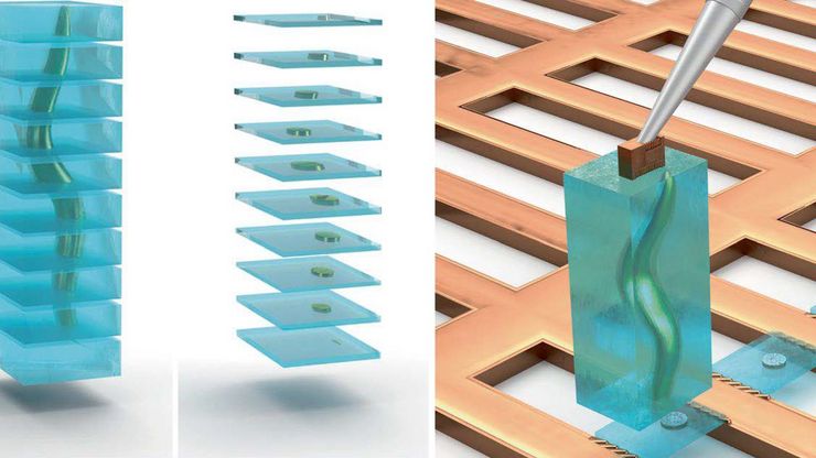
Serial Lift-Out workflow for cryo-ET lamella preparation - Schematic illustration of the Serial Lift-Out method applied to a vitrified biological sample (e.g., C. elegans larva) embedded in bulk vitreous ice (blue). Image courtesy: Prof. Dr. Jürgen M. Plitzko - doi.org/10.1038/s41592-023-02113-5.

Cryo-ET Sample Preparation: From Waffle Method to Serial Lift-Out

Cryo-ET sample preparation becomes more demanding when specimens are thicker, larger, or more complex. This webinar brings together four perspectives on how high-pressure freezing can be connected…
Waffle method lamella preparation and cryo-ET results - Representative workflow and results of lamella preparation from a vitrified C. elegans L1 larva using the waffle method. Image courtesy: Prof. Dr. Jürgen M. Plitzko - doi.org/10.1038/s41592-023-02113-5-

Waffle Method Workflow: From HPF to Cryo-ET Lamellae

Waffle freezing provides an HPF-based route to cryo-ET sample preparation. This workflow guide follows the process from grid and carrier assembly to vitrification, cryo-FIB milling, lamella…
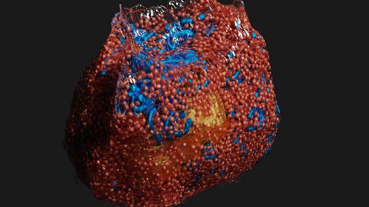
3D rendering of a mouse beta cell with mitochondria (blue), insulin SGs (orange), microtubules (red), nucleus (yellow), and plasma membrane (transparent).

High-Pressure Freezing Protocols for Ultrastructural 3D EM

High pressure freezing (HPF) can help preserve hydrated cells and tissues close to their biological state at the moment of immobilization, supporting more reliable ultrastructural interpretation than…
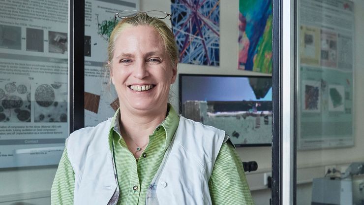
Claudia Mayrhofer ZFE Graz

Ultramicrotome UC Enuity in Practice: Stable 15 nm Sections at ZFE

After using the UCT and UC6 ultramicrotomes, Claudia Mayrhofer calls UC Enuity a leap in stability—so robust that vibrations and temperature shifts don’t spoil sections, even with multiple users. Auto…
Sectioning resin-embedded sambles

超薄切片技术电子书:定位、修块& 对刀

超薄切片技术正经历日新月异的发展,当今的显微镜系统对高质量切片、精准定位以及可重复的工作流程提出了更高要求。这本电子书整合了专家应用指南、自动化方法及实操指导,旨在帮助从初学者到资深镜检人员的每一位用户,在电镜、光电联用及体电镜工作流程中,获得一致且可靠的超薄切片。
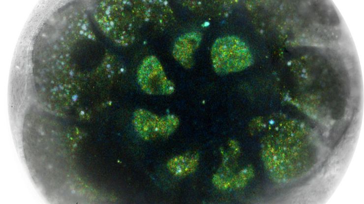
Foraminifera (Ammonia confertitesta) labeled with membrane-permeable calcein, high-pressure frozen in salt water using EM ICE. The sample was cryo-planed and targeted with the M205 on the Cryo-Fluo Enuity, then transferred under cryo conditions to the Cryo-Stellaris for widefield and confocal imaging, revealing details of the staining pattern. Image courtesy: David Evans, University of Southampton.

High-Pressure Freezing for Organoids: Cryo CLEM & FIB Lift Out

Master cryo EM workflow steps for challenging 3D samples: when to choose HPF vs. plunge freezing, reproducible blotting/ice control, contamination aware transfers, Cryo CLEM 3D targeting in organoids,…
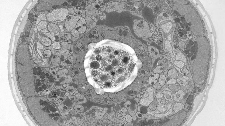
Electron microscope (EM) image of a cross section of C. elegans (roundworm). Courtesy of T. Müller-Reichert, MPI-CBG, Dresden, Germany and K. McDonald, University of California, Berkeley, USA.

高压冷冻简介

水是细胞最主要的组成部分,因此对于维持细胞超微结构至关重要。目前,冷冻固定是固定细胞成分,而不导致其显著结构变化的唯一途径。现阶段有两种常见的方法:投入冷冻与高压冷冻固定。
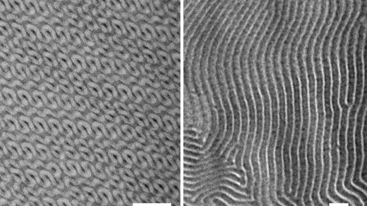
TEM micrographs of polymer sections. Left: Poly(styrene)-b-poly(isoprene). Right: Poly(styrene)-b-poly(methyl methacrylate).

聚合物透射电镜分析用超薄切片技术

本文全面展示了徕卡UC Enuity超薄切片机在聚合物样品超薄切片制备中的优异表现,无论是常温还是低温环境,它都能提供理想的分析样本。文中展示的高分辨率二维及三维TEM图像,有力印证了该仪器在聚合物结构分析领域,对于获得精确、可重复的样品制备结果不可或缺。
Scroll to top