联系我们

James DeRose , Ph.D.

James DeRose

James DeRose is a Global Marketing Communication Manager at Leica Microsystems. His focus is on optimized content creation for applications concerning life science, material science, as well as industry and manufacturing. He is involved with application notes, case studies, technical reports, white papers, application pages, product pages, social media posts, emails, testimonials, and other materials. He has worked at Leica Microsystems since 2013. In the past, he worked on applications development projects in the fields of interfacial chemistry and physics, thermal and chemical engineering, corrosion and metallography, surface coatings, materials science, biotechnology, and cellular biology. He has expertise with various types of microscopy and analytical methods.

Automated Laser Microdissection for Proteome Analysis

组织中的精密空间蛋白质组学信息

尽管可使用基于成像和质谱的方法进行空间蛋白质组学研究,但是图像与单细胞分辨率蛋白丰度测量值的关联仍然是个巨大的挑战。最近引入的一种方法,深层视觉蛋白质组学(DVP),将细胞表型的人工智能图像分析与自动化的单细胞或单核激光显微切割及超高灵敏度的质谱分析结合在了一起。DVP在保留空间背景的同时,将蛋白丰度与复杂的细胞或亚细胞表型关联在一起。
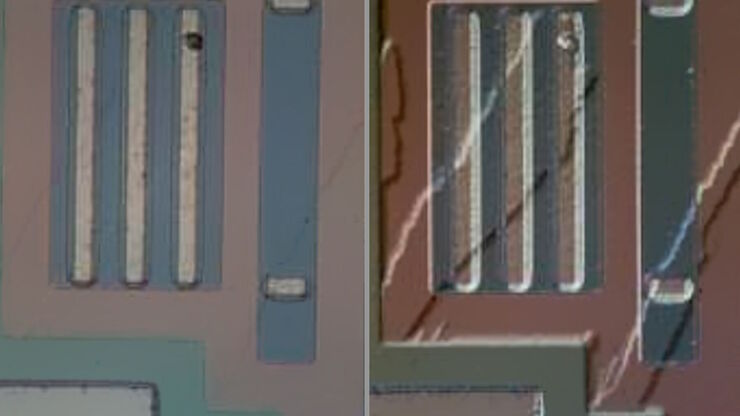
[Translate to chinese:] These images show the microstructure of a hard metal with 10% cobalt which is used for heavy-duty tools. The large increase in magnification of the right image (compared to the left) has a risk of being outside the useful range or, in other words, empty magnification.

当心“无效”放大率

在一个最简化的情况,光学显微镜由一个靠近标本的透镜(物镜)和一个靠近眼睛的透镜(目镜)组成。显微镜放大率是两个显微镜透镜系数的乘积。比如,40倍物镜和10倍目镜可以得到400倍放大率。
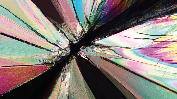
Image of a tartaric-acid crystal taken with polarization microscopy. Tartaric acid, a diprotic, aldaric carboxylic acid, is a naturally occurring organic compound notably found in grapes.

偏振光显微观察

偏光显微镜通常应用于材料科学和地质学领域,根据矿物的折射特性和颜色来识别矿物。在生物学中,偏光显微镜通常用于晶体等双折射结构的识别或成像,或用于植物细胞壁中纤维素和淀粉粒的成像。
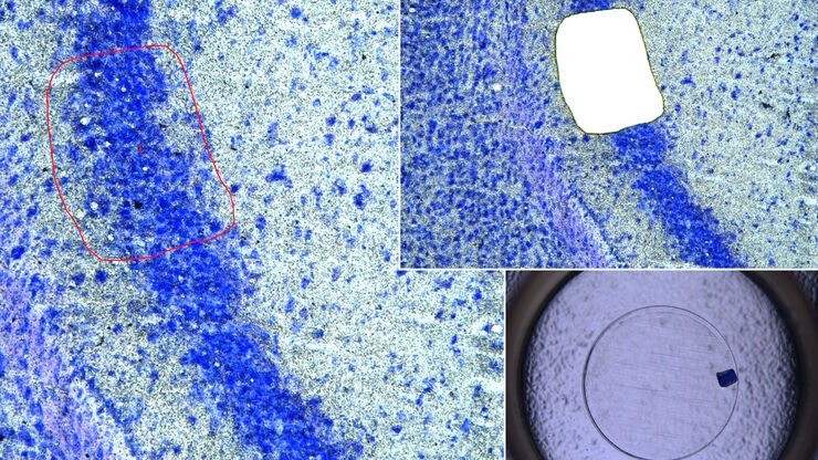
[Translate to chinese:] Mouse brain (left) microdissected with a 10x objective (upper right). Inspection of the collection device (lower right).

激光微切割(LMD)促进的分子生物学分析

使用激光微切割(LMD)提取生物分子、蛋白质、核酸、脂质和染色体,以及提取和操作细胞和组织,可以深入了解基因和蛋白质的功能。它在神经生物学、免疫学、发育生物学、细胞生物学和法医学等多个领域有广泛应用,例如癌症和疾病研究、基因改造、分子病理学和生物学。LMD 也有助于研究蛋白质功能、分子机制及其在转导途径中的相互作用。
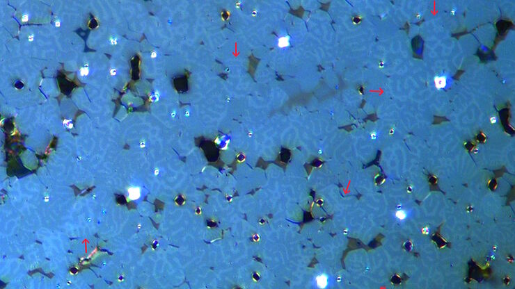
[Translate to chinese:] Image of magnetic steel taken with a 100x objective using Kerr microscopy. The magnetic domains in the grains appear in the image with lighter and darker patterns. A few domains are marked with red arrows. Courtesy of Florian Lang-Melzian, Robert Bosch GmbH, Germany.

使用克尔显微镜快速可视化钢中的磁畴

磁性材料中磁畴与偏振光相互作用后光的旋转,称为克尔效应,使得使用克尔显微镜对磁化样品进行研究成为可能。它可以快速可视化材料表面的磁域。对于用于电气和电子设备的磁性材料(例如钢合金)的高效研发和质量控制,克尔显微镜可以发挥重要作用。本文详细描述了如何使用克尔显微镜对钢合金晶粒中的磁域进行成像。
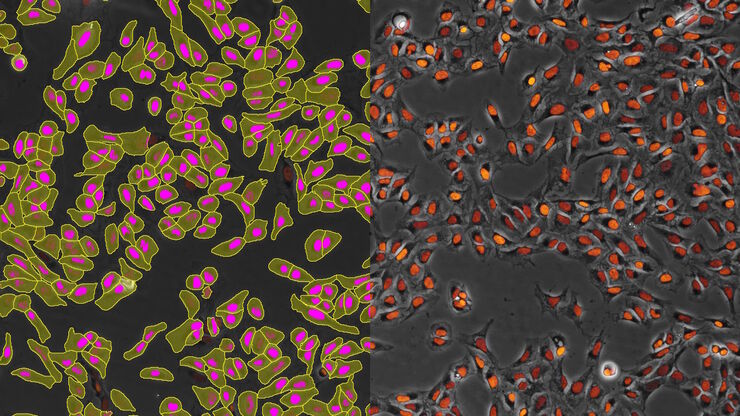
[Translate to chinese:] AI-based cell counting performed with a phase-contrast and fluorescence image using the Mateo FL microscope.

利用AI增强的细胞计数实现精准和高效

本文描述了利用AI进行精确和高效的细胞计数。准确的细胞计数对于 2D 细胞培养的研究至关重要,例如细胞动力学、药物发现和疾病建模。精确的细胞计数对于确定细胞存活率、增殖速率和实验条件的影响至关重要。这些因素对于可靠和稳健的结果至关重要。描述了基于人工智能的方法如何显著提高细胞计数的准确性和速度,从而对细胞研究产生重大影响。
[Translate to chinese:] AI-based transfection analysis (left) of U2OS cells which were transfected with a fluorescently labelled protein. A fluorescence image of the cells (right) is also shown. The analysis and imaging were performed with Mateo FL.

利用AI实现细胞转染的高效分析

本文探讨了AI(AI)在优化 2D 细胞培养研究中转染效率测量中的关键作用。对于理解细胞机制而言,精确可靠的 2D 细胞培养转染效率测量至关重要。靶向蛋白的高转染效率对于包括活细胞成像和蛋白纯化在内的实验至关重要。手动估计存在不一致性和不可靠性。借助AI的力量,可以实现高效可靠的转染研究。
[Translate to chinese:] Image of confluent cells taken with phase contrast (left) and analyzed for confluency using AI (right).

通过 AI 汇合度提高 2D 细胞培养的精度

本文解释了如何利用人工智能(AI)进行高效、精确的 2D 细胞培养汇合度评估。准确评估细胞培养的汇合度,即表面积覆盖的百分比,对于可靠的细胞研究至关重要。传统方法使用视觉检查或简单算法,使结果不客观和精确,尤其是对于用于药物发现、组织工程和再生医学的复杂细胞系。利用自动化图像分析和深度学习算法的方法提供更好的精度,并可以增强实验结果。
[Translate to chinese:] Image of murine dopaminergic neurons which have been marked for laser microdissection (LMD).

利用激光显微切割(LMD)在空间背景下分离神经元

在阿尔茨海默病之后,帕金森病是第二常见的进行性神经退行性疾病。在首发症状出现之前,中脑中高达70%的多巴胺释放神经元已经死亡。本文描述了如何使用现代激光显微切割(LMD)方法帮助解决帕金森病之谜。研究涉及在空间背景下分离和分析神经元。这些细胞来自帕金森病患者的死后黑质组织样本,以便深入了解该病的分子机制。
Region of a patterned wafer inspected using optical microscopy and automated and reproducible DIC (differential interference contrast). With DIC users are able to visualize small height differences on the wafer surface more easily.

6英寸晶圆检测显微镜:可靠观察细微高度差异

本文介绍了一种配备自动化和可重复的DIC(微分干涉对比)成像的6英寸晶圆检测显微镜,无论用户的技能水平如何。制造集成电路(IC)芯片和半导体组件需要进行晶圆检测,以验证是否存在影响性能的缺陷。这种检测通常使用光学显微镜进行质量控制、故障分析和研发。为了有效地可视化晶圆上结构之间的小高度差异,可以使用DIC。
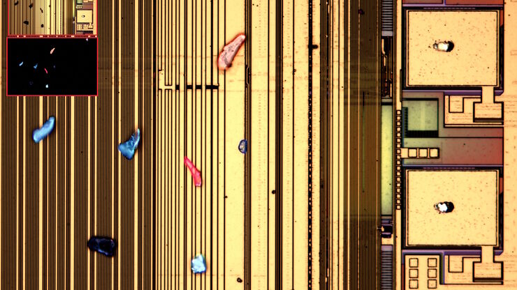
[Translate to chinese:] Optical microscope image, which is a composition of both brightfield and fluorescence illumination, showing organic contamination on a wafer surface. The inset images in the upper left corner show the brightfield image (above) and fluorescence image (below with dark background).

晶圆上的光刻胶残留和有机污染物的可视化

随着半导体上集成电路(IC)的尺寸低于10纳米,在晶圆检测中有效检测光刻胶残留等有机污染物和缺陷变得越来越重要。光学显微镜仍然是常见的检测方法,但对于有机污染物,明场和其他类型的照明可能会存在局限性。本文讨论了荧光显微镜如何在半导体行业的QC、故障分析和研发过程中有效检测晶圆上的光刻胶残留和其他有机污染物。
[Translate to chinese:] Image of burrs (red arrows) at the edge of a battery electrode acquired with a DVM6 digital microscope.

电池制造过程中的毛刺检测

毛刺是电池电极片边缘可能出现的缺陷,例如在制造过程中的分切环节。它们可能会因诸如短路等故障导致电池性能下降,并引发安全和可靠性问题。毛刺检测是电池生产质量控制的重要部分,对于生产具有可靠性能和寿命的电池至关重要。通过适当照明的光学显微镜可以在生产过程的关键步骤中快速可靠地对电极上的毛刺进行视觉检测。
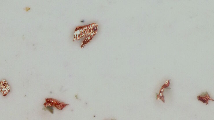
[Translate to chinese:] Particles observed on the surface of a particle trap which could be used for technical cleanliness during battery production.

生产过程中电池颗粒检测

本文介绍了如何利用光学显微镜快速、可靠且经济有效地进行电池颗粒检测与分析。
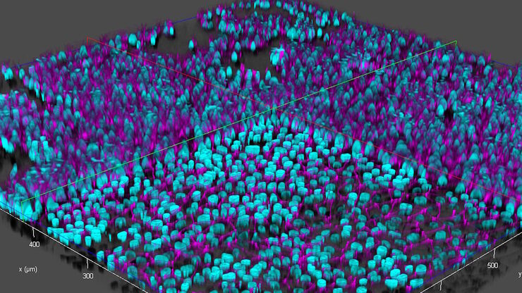
[Translate to chinese:] THUNDER image of brain-capillary endothelial-like cells derived from human iPSCs (induced pluripotent stem cells) where cyan indicates nuclei and magenta tight junctions.

利用多孔板中的细胞培养支撑物对干细胞进行动态研究

本文展示了如何使用THUNDER成像仪高效地对设置在多孔板中的细胞支撑物上的活细胞进行成像,以检查细胞生长情况。
[Translate to chinese:] Particulate contamination in between moving metal plates.

高效清洁度分析的关键因素

在汽车和电子行业,零部件上细小的污染颗粒物也可能影响产品的性能,导致产品出现故障,或使用寿命缩短。对于汽车来说,过滤系统很容易受到影响。对于电子产品来说,印刷电路板(PCB)或连接器上的污染可能会导致短路。因此,清洁度在现代制造业的质量控制中占有核心地位,特别是使用由不同供应商生产的部件时,更要重点关注清洁情况。车辆或设备的关键部件如果受到污染,整个系统就可能发生故障。因此,高效清洁度分析过程必须…
[Translate to chinese:] Microscope equipped with a K7 color CMOS camera for life-science and industry imaging applications.

数码显微镜摄像头和图像分析的基础技术术语定义

现今绝大多数显微镜都配置了摄像头。摄像头的特征通常决定了所采集到的图像是否能够揭示出研究人员希望观察到的现象。但深入到摄像头术语时,技术术语十分繁杂。我们汇总整理了最为重要的术语及其简明释意以便提供方向。这些术语按字母顺序排列。
[Translate to chinese:] Images of the same area of a processed wafer taken with standard (left) and oblique (right) brightfield illumination using a Leica compound microscope. The defect on the wafer surface is clearly more visible with oblique illumination.

显微镜不同观察方法下的快速半导体检测

已刻蚀的晶圆和集成电路(IC)在生产过程中的半导体检测对于识别和减少缺陷非常重要。
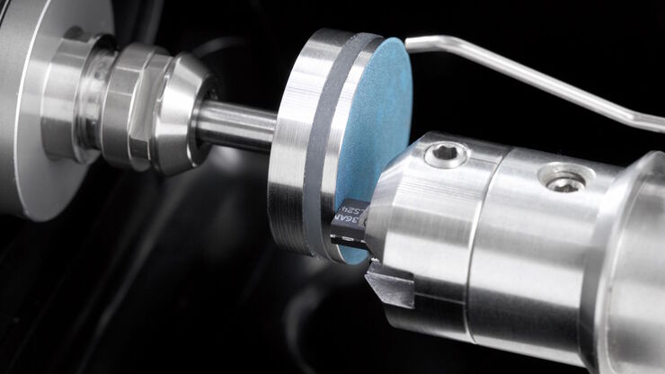
[Translate to chinese:] Preparation of an IC-chip cross section: grinding and polishing of the chip cross section.

电子产品制造截面分析

本文将讨论印刷电路板 (PCB) 和总成 (PCBA)、集成电路 (IC) 和电池组件的横截面为什么对质量控制 (QC)、故障分析 (FA) 和研发 (R&D) 有效,以及如何制备这些横截面。
Image of immunofluorescently labelled cells where mitochondria are indicated with red, nuclei with blue, and actin with green.

采用徕卡THUNDER-DM6B观察SARS-CoV-2感染宿主细胞及其复制过程

冠状病毒2致重度急性呼吸综合征(SARS-CoV-2) 冠状病毒2致重度急性呼吸综合征(SARS-CoV-2)出现于2019年末,并快速传播全世界。由于其大面积的影响,研究人员对病毒的性质进行了深入的研究以期最终阻止大流行。一个重要的方面是病毒如何在宿主细胞中复制。Ogando及其同事的研究已经揭示了SARS-CoV-2的复制动力学、适应能力和细胞病理学。他们的工具之一是用荧光显微镜观察SARS…
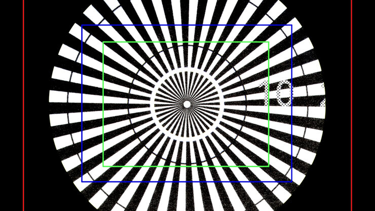
Image of a Siemens star, where the diameter of the 1st black line circle is 10 mm and the 2nd is 20 mm, taken via an eyepiece of a M205 A stereo microscope. The rectangles represent the field of view (FOV) of a Leica digital camera when installed with various C-mounts (red 0.32x, blue 0.5x, green 0.63x).

30000:1放大率到底意味着什么?

关于光学显微镜性能的一个重要标准是放大率。本报告将为数字显微镜用户提供有用的指南,以确定放大率值的有用范围。
Fluorescence microscopy image of liver tissue where DNA in the nuclei are stained with Feulgen-pararosanilin and visualized with transmitted green light.

Epi-Illumination Fluorescence and Reflection-Contrast Microscopy

This article discusses the development of epi-illumination and reflection contrast for fluorescence microscopy concerning life-science applications. Much was done by the Ploem research group…
[Translate to chinese:] Image of an onion flake taken with a basic Leica compound microscope after it was tested for resistance to fungus and mold growth following part 11 of the ISO 9022 standard.

ISO 9022 标准第 11 部分 - 在苛刻条件下测试显微镜

显微镜和其他光学仪器会受到环境因素的影响。环境因素取决于地理位置和使用地点的条件。仪器的坚固性可以通过完善的加速测试方法进行评估。ISO 9022 标准第 11 部分规定了测试光学仪器抗霉菌和真菌生长的方法。
EBSD grain size distribution of the cross section of a gold wire within a silicon matrix from inside a CPU (central processing unit of a computer). The grains are highlighted with arbitrary colors.

高质量EBSD样品制备

本文介绍了一种使用宽离子束研磨技术为“混合”晶体材料制备可靠且有效的EBSD(电子背散射衍射)样品的方法。该方法产生的横截面具有高质量表面,这对于EBSD分析至关重要。电子背散射衍射(EBSD)材料分析是通过扫描电子显微镜(SEM)进行的。制备混合材料(CPU或铝(Al)、金刚石和石墨(C)的复合材料)的横截面,使其具有适合EBSD分析的高质量表面,可能是一个挑战。
Stereo microscopes are often considered the workhorses of laboratories and production sites.

考虑采购体视显微镜时的关键因素

体视显微镜通常是实验室或生产现场“主力”。用户需要花费数小时通过目镜来检查、观察、记录或解剖样本。仔细评估哪些相关应用需要用到体视显微镜,是确保长期满意使用的关键所在。决策者们需要确保自己能够完全依照自己的需求来定制仪器。为帮助用户能更好的选择适合自己的体视镜,本文介绍了几个主要考虑的因素。

利用数码显微镜快速、可靠地对印刷电路板(PCB)及其总成(PCBA)进行显微观察

本文阐释了徕卡数码显微镜DVM6的性能优势,例如简单直观的操作系统、快速简单的放大倍率切换方式,并且可以通过编码准确调取参数。

荧光入门介绍

荧光是George Gabriel Stokes于1852年首次报道的一种现象。他观察到萤石在紫外线照射后开始发光。荧光是光致发光的一种形式,是指一种材料被光照射后会发射出光子。发射光的波长比激发光更长。这种效应又称为斯托克斯位移。
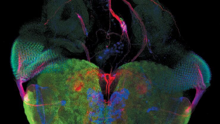
[Translate to chinese:] Raw widefield and THUNDER image of calcium transients in Drosophila embryos. Courtesy A. Carreira-Rosario, Clandinin laboratory, California, USA.

Central Nervous System (CNS) Development and Activity in Organisms

This article shows how studying central nervous system (CNS) development in Drosophila-melanogaster embryos expressing a GCaMP calcium indicator in the neurons can be improved with a THUNDER Imager.
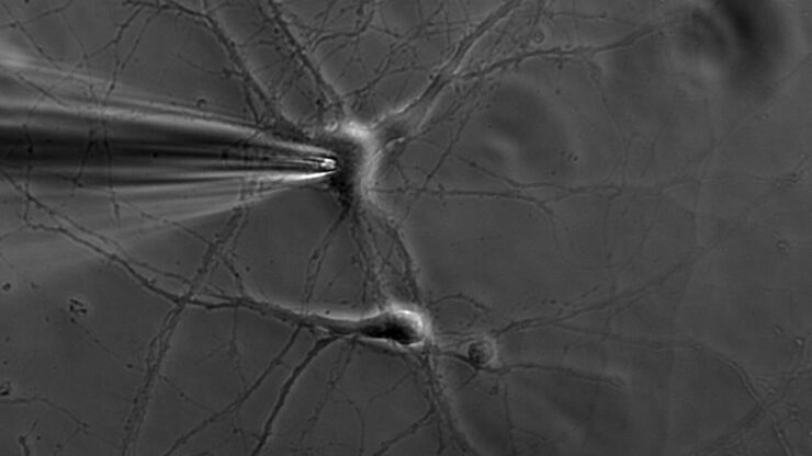
Patch pipette touching a murine hippocampal neuron. Image courtesy of A. Aguado, Ruhr University Bochum, Germany.

什么是膜片钳技术?

离子通道的生理学一直是神经科学家感兴趣的一个重要话题。诞生于1970年代的膜片钳技术开启了电生理学家的新时代。它不仅可以对整个细胞进行高分辨率电流记录,还可以对切下的细胞膜片进行高分辨率电流记录。甚至可以研究单通道事件。然而,由于需要复杂且高灵敏的设备,广泛的生物学背景和高水平的实验技能,电生理学仍然是最具挑战性的实验室方法之一。
[Translate to chinese:] Neurons imaged with DIC contrast.

微分干涉对比(DIC)显微镜

本文阐释了在使用显微镜对未染色透明生物样本进行成像时,微分干涉对比法(DIC)为何是明场照明的绝佳方案。
[Translate to chinese:] The Emspira 3 digital microscope offers what users need for comprehensive visual inspection, including comparison, measurement, and documentation sharing.

数码检测显微镜的工业应用

本文讨论了在选择用于显微分析和质量控制(QC)以及故障分析(FA)和研发(R&D)的数码显微镜之前,用户应当考虑的因素。关键在于需要事先充分了解汽车、电子、机械工程和医疗设备等行业的应用要求和用户需求。显微镜解决方案不仅应当帮助用户实现高效、可靠的显微分析、QC、FA以及研发工作,还应当易于操作、满足用户需求,同时方便报告并分享结果。
Scroll to top