


Advancing ADC Development with Imaging of 3D Tumor Models
Antibody-drug conjugates (ADCs) are targeted therapies designed to selectively deliver drugs to cancer cells. Evaluating ADC behavior in 3D tumor models more closely reflects the physiological…
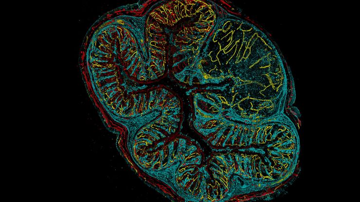
Visualizing Life in 3D at High Speed with Viventis SCAPE
Discover how oblique plane microscopy (OPM) enables light sheet imaging through a single objective, combining illumination and detection in a geometry that is compatible with conventional sample…
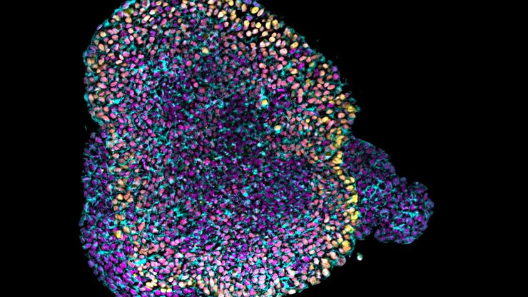
Volumetric Imaging of Cleared Organoids with Light Sheet
Cleared organoid imaging with Viventis Deep light sheet: rapid, high-resolution imaging of large organoids throughout their full volume.
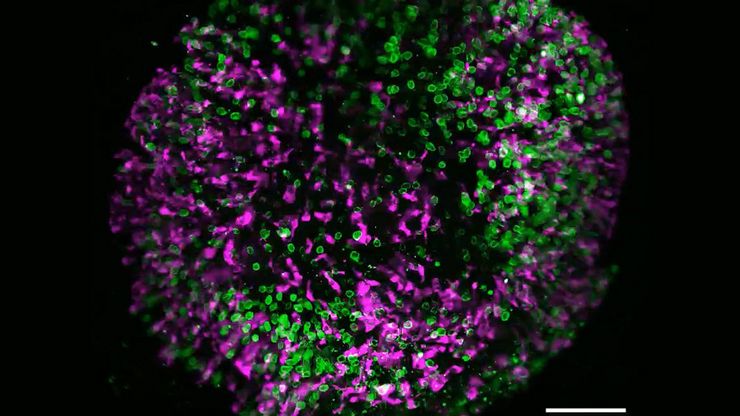
如何深入了解类器官和细胞球模型
在本电子书中,您将了解3D细胞培养模型(如类器官和细胞球)成像的关键注意事项。探索创新型显微镜解决方案,来实时记录类器官和细胞球的动态成像过程。

Fast, High-Contrast Widefield Imaging of Optically Challenging Samples
Live‑cell imaging of large, complex biological samples often requires large fields of view, sub-cellular resolution, high-sensitivity, and fast acquisition – all while maintaining low illumination…
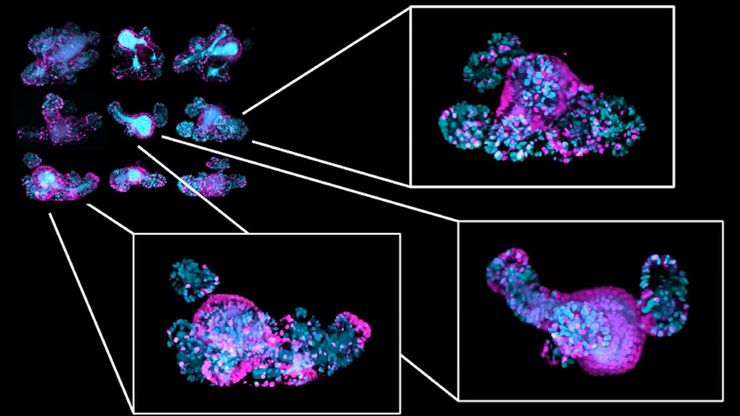
Multiscale Imaging of Organoids: High Content to Light Sheet
Learn multiscale organoid imaging: fixed high content phenotyping, gentle dual view light sheet, and reproducible pipelines that turn 3D data into insights.
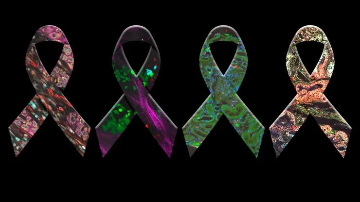
癌症研究中显微镜的历史、发展和趋势
癌症是一种全球性疾病,2020 年全球将新增 1800 万确诊病例,1000 万人死于癌症。预计到 2040 年,病例数将增加约 55%。研究了解癌症的发生、发展以及开发新的诊断工具和治疗方法至关重要。
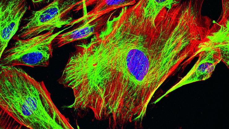
荧光染料应用和特性概述
本文将介绍常用的荧光染料并概述其特性。荧光显微镜借助荧光染料、荧光蛋白或使用抗体的免疫荧光染色来研究特定的细胞成分。由于荧光剂种类繁多,荧光显微镜可用于检查蛋白质、核酸、聚糖、细胞器和其他细胞结构。
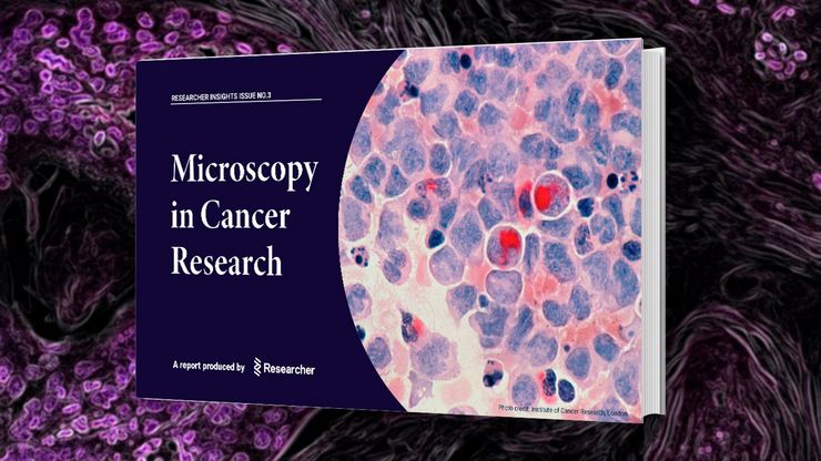
Researchers Insights: Microscopy in Cancer Research
Discover how imaging techniques are driving cancer research forward. In this issue, we present comprehensive multimodal studies using microscopy, as well as new directions in intraoperative cancer…


