
癌症研究中显微镜的历史、发展和趋势
癌症是一种全球性疾病,2020 年全球将新增 1800 万确诊病例,1000 万人死于癌症。预计到 2040 年,病例数将增加约 55%。研究了解癌症的发生、发展以及开发新的诊断工具和治疗方法至关重要。
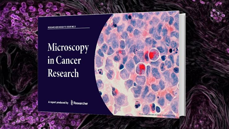
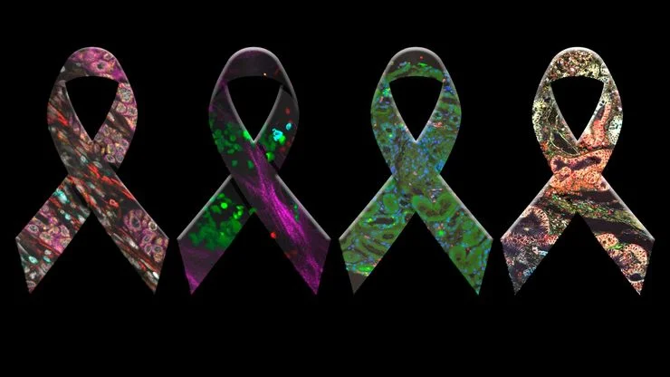
Researchers Insights: Microscopy in Cancer Research
Discover how imaging techniques are driving cancer research forward. In this issue, we present comprehensive multimodal studies using microscopy, as well as new directions in intraoperative cancer…
, Tropomyosin (cardiomyocytes, red) and GFP (primordial cardiac layer, green).")
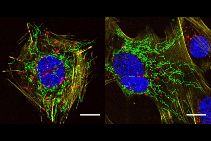
显微镜中的荧光
荧光显微技术是一种特殊的光学显微镜技术。它利用的是荧光色素在一定波长的光激发下发光的能力。通过抗体染色或荧光蛋白标记,可以用这种荧光色素标记感兴趣的蛋白质。这样就可以确定单分子物种的分布、数量及其在细胞内的定位。此外,还可以进行共定位和相互作用研究,使用可逆结合染料(如 Ca2+ 和 fura-2)观察离子浓度,以及观察细胞的内吞和外吞过程。如今,利用荧光显微镜甚至可以对亚分辨率颗粒进行成像。

超分辨率显微镜图片库
由于光的衍射极限,传统共聚焦显微镜无法分辨约240纳米以下的结构。当需要提高分辨率以研究衍射极限尺度以下的结构和分子事件时,会使用超分辨率显微镜技术,如STED、PALM或STORM,或某些解卷积处理方法。

细胞活成像的纳米级扩展
新的STED显微技术方法——TauSTED Xtend,使得在纳米级别下对活体完整样本进行扩展多色成像成为可能。通过结合空间和寿命信息,TauSTED Xtend提供了额外一层信息,允许在极低的光剂量下分辨小细节并在整体结构中解析它们。
, actin network (ATTO 647N), and nuclear pore basket (CF 680R).")
STED样品制备指南
这份指南旨在帮助用户优化受激发射损耗(STED)纳米成像的样品制备,特别是在使用徕卡微系统的STED显微镜时。它提供了单色STED成像用荧光标记的概述,并对其性能进行了评级。

采用徕卡THUNDER-DM6B观察SARS-CoV-2感染宿主细胞及其复制过程
冠状病毒2致重度急性呼吸综合征(SARS-CoV-2)
冠状病毒2致重度急性呼吸综合征(SARS-CoV-2)出现于2019年末,并快速传播全世界。由于其大面积的影响,研究人员对病毒的性质进行了深入的研究以期最终阻止大流行。一个重要的方面是病毒如何在宿主细胞中复制。Ogando及其同事的研究已经揭示了SARS-CoV-2的复制动力学、适应能力和细胞病理学。他们的工具之一是用荧光显微镜观察SARS…

采用单损耗激光的五色FLIM-STED显微镜
网络研讨会,内容涉及使用单一损耗激光和荧光寿命phasor分离技术的五色STED技术。
A Versatile Palette of Fluorescent Probes
Researchers at the Max Planck Institute for Medical Research in Heidelberg have developed a general strategy to synthesize live-cell compatible fluorogenic probes, and the result are the new MaP (Max…
