Filter articles
标签
产品

Loading...
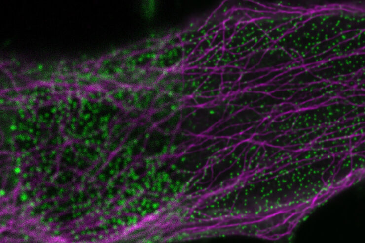
使用深度学习技术追踪单细胞
人工智能解决方案在显微镜领域的应用不断拓展。从自动化目标分类到虚拟染色,机器学习和深度学习技术在帮助显微镜学家简化分析工作的同时,也在持续推动科学技术领域的突破。
Loading...
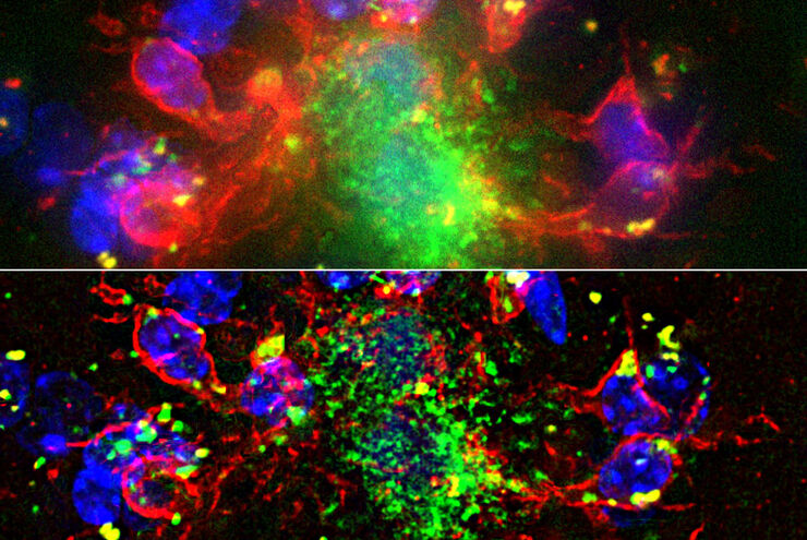
Computational Clearing - 增强3D标本成像
本次网络研讨会旨在阐明有助于THUNDER显微成像仪实现三维样品可视化的关键规格,并改进研究人员的成像相关工作流程。
Loading...
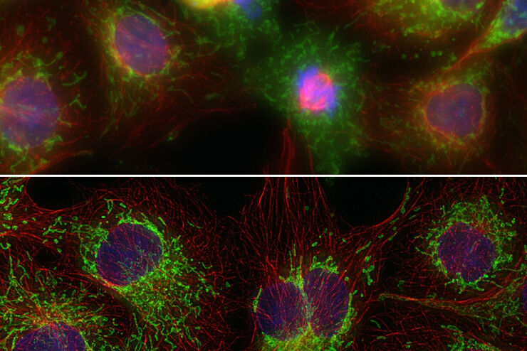
THUNDER成像:高效、灵活、易操作,让您的日常成像工作流更轻松
本次网络研讨会将展示 THUNDER 在许多不同生命科学应用中的多功能性和性能:从计数视网膜切片中的细胞核和癌组织切片中的 RNA 分子,到监测阿拉伯芥幼苗中的钙波等等。
Loading...
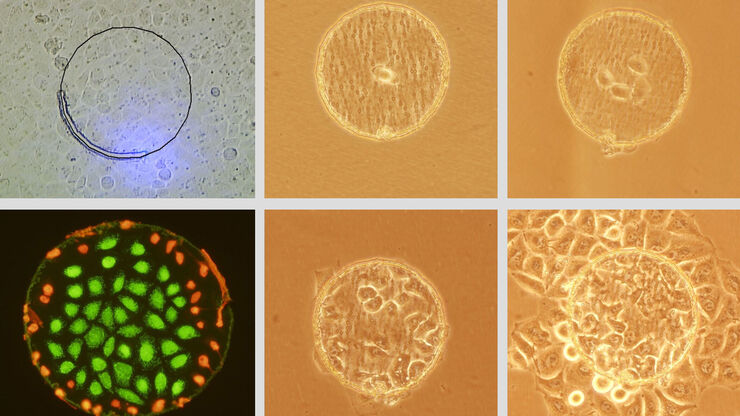
Live Cell Isolation by Laser Microdissection
Laser microdissection is a tool for the isolation of homogenous cell populations from their native niches in tissues to downstream molecular assays. Beside its routine use for fixed tissue sections,…
