


Advancing ADC Development with Imaging of 3D Tumor Models
Antibody-drug conjugates (ADCs) are targeted therapies designed to selectively deliver drugs to cancer cells. Evaluating ADC behavior in 3D tumor models more closely reflects the physiological…
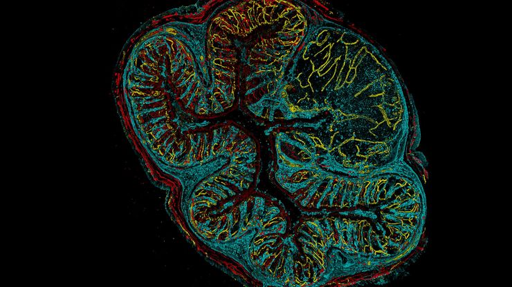
Visualizing Life in 3D at High Speed with Viventis SCAPE
Discover how oblique plane microscopy (OPM) enables light sheet imaging through a single objective, combining illumination and detection in a geometry that is compatible with conventional sample…
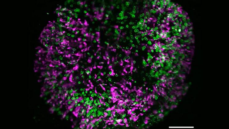
如何深入了解类器官和细胞球模型
在本电子书中,您将了解3D细胞培养模型(如类器官和细胞球)成像的关键注意事项。探索创新型显微镜解决方案,来实时记录类器官和细胞球的动态成像过程。
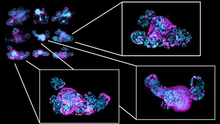
Multiscale Imaging of Organoids: High Content to Light Sheet
Learn multiscale organoid imaging: fixed high content phenotyping, gentle dual view light sheet, and reproducible pipelines that turn 3D data into insights.

比例成像
细胞的许多基本功能在很大程度上依赖于离子(例如钙、镁)、电压势和细胞质与周围细胞外空间之间的 pH 值的微妙但动态的平衡。这些平衡的变化会显著改变细胞的行为和功能。因此,实时测量细胞内离子、电压和 pH…

癌症研究中显微镜的历史、发展和趋势
癌症是一种全球性疾病,2020 年全球将新增 1800 万确诊病例,1000 万人死于癌症。预计到 2040 年,病例数将增加约 55%。研究了解癌症的发生、发展以及开发新的诊断工具和治疗方法至关重要。
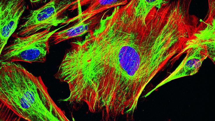
荧光染料应用和特性概述
本文将介绍常用的荧光染料并概述其特性。荧光显微镜借助荧光染料、荧光蛋白或使用抗体的免疫荧光染色来研究特定的细胞成分。由于荧光剂种类繁多,荧光显微镜可用于检查蛋白质、核酸、聚糖、细胞器和其他细胞结构。
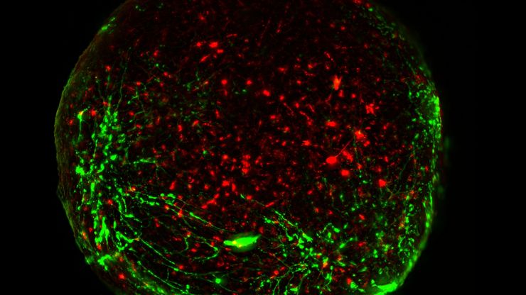
活细胞成像指南
在生命科学各研究领域的广泛应用中,活细胞成像是一种不可或缺的工具,用于观察细胞在尽可能接近活体(即活的、活跃的)状态下的情况。本指南回顾了确保成功进行活细胞成像的各种重要注意事项,并介绍了各种旨在克服常见挑战的高性能解决方案。这些进展使我们能够对细胞生理学和动力学有新的认识。
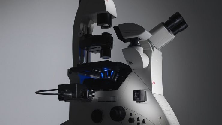
选择研究用显微镜时应考虑的因素
光学显微镜通常是生命科学研究实验室的核心设备之一。它可用于各种应用,揭示许多科学问题。因此,显微镜的配置和功能对其应用范围至关重要,从明视野显微镜到荧光显微镜,再到活细胞成像。本文简要概述了显微镜的相关功能,并总结了在选择研究用显微镜时应考虑的关键问题。


