Aivia
显微镜软件
产品
首页
Leica Microsystems
Aivia AI 图像分析软件
借助人工智能,更快、更轻松地获得洞察力
阅读我们的最新文章
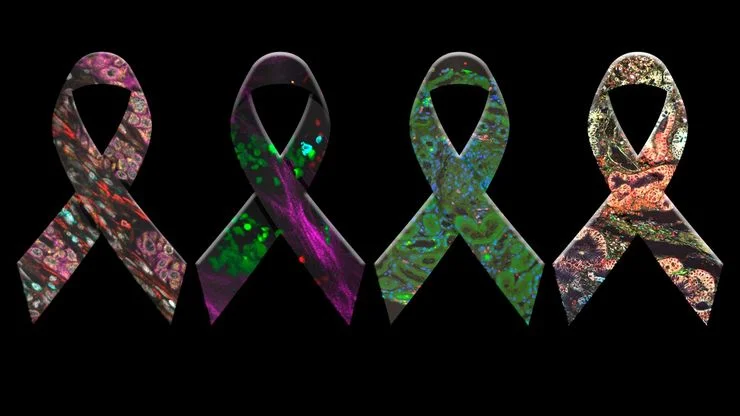
癌症研究中显微镜的历史、发展和趋势
癌症是一种全球性疾病,2020 年全球将新增 1800 万确诊病例,1000 万人死于癌症。预计到 2040 年,病例数将增加约 55%。研究了解癌症的发生、发展以及开发新的诊断工具和治疗方法至关重要。
 and astrocytes (green) in a cortical spheroid derived from human induced pluripotent stem cells.")
活细胞成像指南
在生命科学各研究领域的广泛应用中,活细胞成像是一种不可或缺的工具,用于观察细胞在尽可能接近活体(即活的、活跃的)状态下的情况。本指南回顾了确保成功进行活细胞成像的各种重要注意事项,并介绍了各种旨在克服常见挑战的高性能解决方案。这些进展使我们能够对细胞生理学和动力学有新的认识。
.")
人工智能驱动的乳腺癌研究多重染色成像空间分析工具
乳腺癌(BC)是女性因癌症死亡的主要原因,研究查肿瘤微环境(TME)对于阐明肿瘤进展机制至关重要。利用超多标染色空间蛋白质组学技术系统地绘制肿瘤微环境图谱可以提高精准免疫肿瘤学的能力。在这里,我们将基于人工智能的高倍空间分析应用于BC组织,研究免疫细胞类型和生物标记物,从而深入了解受免疫疗法反应的TME分子机制。
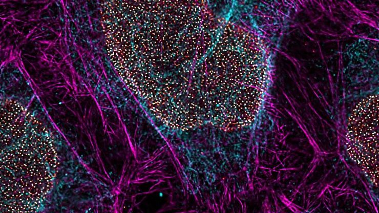
 and tubulin (magenta), acquired using Viventis Deep. Courtesy of Akanksha Jain, Treutlein Lab ETH-DBSSE Basel (Switzerland).")
如何深入了解类器官和细胞球模型
在本电子书中,您将了解3D细胞培养模型(如类器官和细胞球)成像的关键注意事项。探索创新型显微镜解决方案,来实时记录类器官和细胞球的动态成像过程。

体电子显微学与人工智能图像分析
该文章详细阐述了利用体电子显微镜技术 (volume-SEM) 结合人工智能辅助图像分析,对生物组织进行三维研究的工作流程。研究的重点是一种名为毛滴虫的原生动物,这是一种有鞭毛的寄生虫,是导致性传播感染——滴虫病的病原体。为了可视化其复杂的内部结构,研究人员采用了体电子显微镜技术,通过对一系列超薄切片进行成像来重建三维模型。
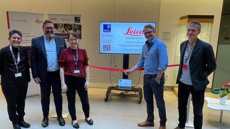
Micron Bioimaging Facility at the University of Oxford
Find out how to access cutting-edge microscopy systems, with dedicated training and support, at the Micron Bioimaging Facility at the University of Oxford’s Department of Biochemistry.

罕见疾病 CRISPR 疗法的开发与风险解除
Fyodor Urnov博士和Sadik Kassim博士最初是在ASGCT 2025会议上作这一按需演讲的,演讲的重点是遗传医学中的一个关键挑战:如何将CRISPR疗法从单一疾病解决方案扩展到平台方法,特别是针对罕见的儿科遗传疾病。Urnov 博士展示了由 Matthew Kan 博士领导的创新基因组研究所的工作,这是 IGI-Danaher Beacon for CRISPR Cures…

用于三维生物成像的集成连续切片与冷冻电镜工作流程
本场网络研讨会探讨了集成化工具如何支持从样品制备到图像分析的电子显微镜全流程。专家Andreia Pinto博士、Adrian Boey博士与Hoyin Lai博士将介绍UC Enuity超薄切片机和Aivia图像分析平台,并演示这些工具如何同时适用于常温与低温实验环境。会议内容包含阵列断层成像、基于深度学习的图像分割、以及生物成像中cryo-lift-out工作流程的实际案例解析。

多重成像揭示结肠癌的肿瘤免疫格局
由于抗药性和复发,癌症免疫疗法获益者寥寥无几,而针对癌症免疫周期多个步骤的组合治疗策略可能会改善治疗效果。这项研究表明,高通量空间蛋白质组学可用于识别细胞生物标志物之间的相互作用,并通过绘制肿瘤免疫微环境图来指导精准的组合疗法。

神经科学研究指南
神经科学通常需要研究具有挑战性的标本,以更好地了解神经系统和疾病。徕卡显微镜帮助神经科学家深入了解神经元功能。
")
如何优化多标成像技术推动3D空间组学发展
本次网络研讨会上,徕卡显微系统的Julia Roberti博士与Luis Alvarez博士将介绍STELLARIS共聚焦平台的全新功能SpectraPlex,该技术可实现超多标三维空间成像。该技术旨在通过实现超多标成像且无需频繁人工干预,从而简化和增强空间生物学应用。

Boston and San Francisco Innovation Hubs
Boston and San Francisco Innovation Hubs are here to help you advance scientific discovery. We provide researchers access to state-of-the-art microscope technology and expert guidance. Located in the…

利用空间蛋白质组学工作流程改革研究工作
空间蛋白质组学是《自然-方法》2024 年度方法,正在推动癌症、免疫学等领域的研究进展。通过将定位数据与组织中蛋白质的高通量成像结合起来,研究人员可以发现疾病进展和治疗反应方面的洞察力,从而更好地了解人类生物学。在这里,您可以了解更多有关空间生物学的信息,以及徕卡显微系统的工具如何推动蛋白质生物标记的可视化和分析取得进展。

利用人工智能图像分析工具更快、更轻松地获得洞察力
了解 Aivia 如何通过快速设置、准确的人工智能检测和简便的批量处理功能,帮助科学家简化图像分析。

揭开类器官模型在生物医学研究中的秘密
准备深入了解类器官和3D培养物的世界,它们是促进我们了解人类健康的重要工具。浏览这些复杂的结构并获取清晰的图像进行分析是一项挑战。在本次活动中,来自牛津大学和伦敦大学学院的研究人员将与我们一起展示Thunder Imager Cell转盘共聚焦系统 如何提供更有说服力的高质量数据,以便深入了解各种模型。
 applied. Image courtesy of Samuel East, Uncommon Bio.")
利用新型可扩展的干细胞培养设计未来
具有远见卓识的生物技术初创企业 Uncommon Bio 正在应对世界上最大的健康挑战之一:食品可持续性。在这次网络研讨会上,干细胞科学家塞缪尔-伊斯特(Samuel East)将展示他们如何使细胞农业的干细胞培养基既安全又经济可行。了解他们如何将培养基成本降低 1000 倍,并开发出不含动物成分、食品安全的 iPSC 培养基。
, microglia (TMEM119, IBA1), and Alzheimer’s-associated markers (β-amyloid and p-Tau217).")
利用大数据探索阿尔茨海默病的空间蛋白组
阿尔茨海默病是一种遗传性和散发性的神经退行性疾病,导致中晚年认知能力下降,特征为β-淀粉样蛋白斑块和 tau蛋白 缠结。由于治疗选择有限,新的研究策略至关重要。Cell DIVE 多重成像解决方案可以对阿尔茨海默病脑组织进行研究,揭示,可能新的研究方向。这里我们展示了 Cell DIVE 多重成像仪的图像查看器,用户能够直接在自己的浏览器中访问完整的阿尔茨海默病多重数据集。

利用大数据查看器揭示结肠癌隐藏的复杂性
结直肠癌是一种的重大健康负担。虽然手术初期有效,但部分患者会发展为预后不良的复发性继发疾病,需要采用免疫疗法等先进治疗手段。利用空间生物学方法,如 Cell DIVE 多重成像技术,可为开发新型治疗方案提供关键洞见。通过 Minerva 图像查看器在浏览器中访问完整的 Cell DIVE 数据集,进一步探索这些发现。

用大数据视角深入了解胰腺癌研究
胰腺癌由于其靠近主要器官难以分辨和难治疗,死亡率接近 40%,。这个研究探讨了胰腺导管腺癌(PDAC)的复杂生物学机制,研究了代谢、凋亡和免疫中肿瘤侵袭性的相关分子结构和空间决定因素。可以访问您的浏览器中的完整 Cell DIVE 数据集,以深入了解这些发现。

利用人工智能驱动的空间蛋白质组学绘制肿瘤免疫图谱
未经治疗肿瘤的空间图谱分析可呈现肿瘤免疫结构的整体特征,有助于理解治疗反应。具有免疫活性的小鼠模型对于识别肿瘤发生发展过程中免疫依赖性事件至关重要。要表征这些具有完整免疫系统及相互作用细胞组分的模型,需要采用多重标记分析技术。我们展示了一种基于人工智能的空间蛋白质组学方法,用于研究小鼠癌组织中的肿瘤-免疫互作机制。

深度视觉蛋白质组学提供精确的空间蛋白质组信息
尽管可使用基于成像和质谱的方法进行空间蛋白质组学研究,但是图像与单细胞分辨率蛋白丰度测量值的关联仍然是个巨大的挑战。最近引入的一种方法,深层视觉蛋白质组学(DVP),将细胞表型的人工智能图像分析与自动化的单细胞或单核激光显微切割及超高灵敏度的质谱分析结合在了一起。DVP在保留空间背景的同时,将蛋白丰度与复杂的细胞或亚细胞表型关联在一起。

阿尔茨海默病神经免疫相互作用的空间分析
阿尔茨海默病(AD)是一种复杂的神经退行性疾病,以神经原纤维缠结、β-淀粉样斑块和神经炎症为特征。这些功能障碍由局部免疫反应触发或加剧。因此,在空间背景下理解神经免疫相互作用对于阐明 AD 发病机制至关重要。本研究采用 Cell DIVE 多重成像技术和 Aivia 人工智能辅助空间分析工具,探究 AD 病理标志物周围免疫细胞的特征。

空间生物学指南
什么是空间生物学?在后组学时代,研究人员如何利用空间生物学工具来满足生物学问题日益增长的需求?本文简要概述了空间生物学及其技术,以及这一快速发展中的领域的关键研究问题。

使用空间多重化探测人类阿尔茨海默病皮层切片
阿尔茨海默病(AD)是最常见的神经退行性疾病,其特征是认知功能的逐渐下降。对 AD 大脑的空间分析可能揭示细胞关系,从而促进对疾病病因的更好理解。本研究捕捉了 AD 皮层组织成分的全球概述,并强调了 Cell DIVE 成像的简化工作流程,从数据采集到使用 Aivia 软件的基于人工智能的分析,最终实现更快的洞察。
.")
您的 3D 类器官成像和分析工作流程效率如何?
类器官模型已经改变了生命科学研究,但优化图像分析协议仍然是一个关键挑战。本次网络研讨会探讨了类器官研究的简化工作流程,首先是实时的三维细胞培养检查,接下来是高速、高分辨率的三维成像,生成清晰的图像和更纯净的数据,以便对生长速率、细胞迁移和三维细胞相互作用等参数进行准确地人工智能分割和量化,从而实现更深入的洞察。

激光显微切割技术如何助力神经科学研究取得开创性进展?
玛尔塔·帕特林尼博士,卡罗林斯卡学院的高级科学家,分享了她在成人人类神经发生开创性研究中使用激光显微切割(LMD)的经验,并提供了关于LMD在空间蛋白质组学和精准医学中未来应用潜力的个人见解。
 tissue.")
基于人工智能的多重图像分析以探索结肠腺癌
在这项研究中,我们展示了一种利用Cell DIVE和AIVIA软件的空间生物学工作流程,以绘制结肠腺癌中的肿瘤免疫景观图。

肿瘤空间微环境的元癌症分析
研究 TME中肿瘤、基质和免疫细胞之间的相互作用需要采用超多重免疫荧光成像方法。在这里,我们分析了一组Cell Signaling Technology(CST®)抗体,这些抗体针对肺癌、结肠癌和胰腺癌等癌症的标志物。通过Cell DIVE成像和Aivia中的聚类分析,我们确定了TME中的空间相互作用,包括组织特异性和共有的相互作用。
 tissue.")
通过成像和AI绘制结直肠癌的景观
结肠癌是一种高负担疾病。尽管进行了化疗干预和手术切除,但疾病可能会复发。了解结肠癌微环境对于改善治疗效果是必要的。在这里,我们使用空间生物学方法,通过Cell DIVE和 Aivia可视化结肠腺癌组织中的30个生物标志物。我们探讨了肿瘤组织的血管化、免疫细胞反应和细胞增殖。

基于 AI 引导的多重二维数据向空间洞察的转化
Aivia 13 能够处理大型二维图像,使研究人员能够通过检测数百万个对象和自动聚类多达 30 个标记物,深入理解其表型周围的微环境。

利用 SPARCS 探索亚细胞空间表型
功能日益强大的显微镜可提供信息丰富的各种细胞表型数据。如果与深度学习的最新进展相结合,这将成为在基因筛选中读出感兴趣的生物表型的理想技术。在本网络讲座中,您将了解到空间分辨 CRISPR 筛选 (SPARCS),这是一种利用自动化高速激光显微切割技术在人类基因组尺度上揭示各种亚细胞空间表型的平台。

借助人工智能,揭示复杂而密集的神经元图像中的洞察
神经元的3D形态学分析通常需要使用不同的成像模式,捕捉多种类型的神经元,并在各种密度下相连的传统Leica SP8显微镜采集多达解神经元的形态,这对许多研究人员来说仍然是一个耗时的挑战。

神经科学显微镜面临哪些挑战?
显微镜是神经科学研究领域的强大工具。不过,当涉及到对神经过程进行成像以及使用不同的样品类型(例如厚神经组织或脑类器官)时,科研人员可能会面临到很多挑战。这本30页的电子书包含众多真实的案例,以讨论我们最常见到的一些挑战,同时展示了如何使用THUNDER 成像技术克服这些挑战。

人工智能显微成像能够高效检测稀有事件
对稀有事件进行定位和选择性成像是许多生物样本研究过程的关键。然而,由于时间限制和高度的复杂性,有些实验无法做到,从而限制了获得新发现的前景。通过基于人工智能的显微成像检测稀有事件,这种工作流程将智能样本导航、图像采集工具和人工智能驱动的图像分析等不同功能融合起来共同协作,能够克服上述局限性。
 stained to show the nucleus")
使用Mica的人工智能显微镜软件进行 3D 空间分析
本期MicaCam为您提供切实的建议,教您从显微镜图像中提取可发表级别的分析结果。本期的特邀嘉宾来自徕卡显微系统的Luciano Lucas,他将为大家展示如何使用MICA的AI赋能软件进行图像分析。他将深度分析两张MICA的3D成像,探究不同可见生物元素之间的空间关系。本期的最后将会介绍如何创作高保真视频动画以及其他可用于发表文章的结果。

精确分析宽视野荧光图像
利用荧光显微镜的特异性,即便是使用厚样品和大尺寸样品,研究人员也能够快速轻松地准确观察和分析生物学过程和结构。然而,离焦荧光会提高背景荧光,降低对比度,影响图像的精确分割。THUNDER 与Aivia 的组合可以有效解决这一问题。前者可以消除图像模糊,后者会使用人工智能技术自动分析宽视野图像,提高操作速度和精确性。下面,我们来详细了解下这一协作方法。

在显微图像分析中运用机器学习技术
显微成像技术最近取得了令人振奋的进展,因此,在生物医学研究中采集的图像数据无论质量还是数量都呈指数级增长。但是,分析日益复杂的大型图像数据集以提取有意义的信息可能是一个既枯燥又耗时的过程,而且容易出现人为误差和偏差,这经常给许多研究人员造成生产效率瓶颈。
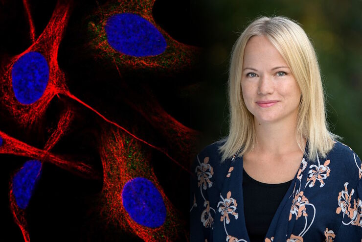
在显微成像和图像分析中运用人工智能和机器学习技术
Emma Lundberg 教授是瑞典 KTH 皇家理工学院细胞生物学蛋白质组学教授。她还是细胞图谱项目的总监,该项目是瑞典人类蛋白质图谱(HPA)项目不可或缺的组成部分,后者是用于研究人类蛋白质组的开源资源。细胞图谱项目是 HPA 的一部分,提供人类细胞系中 RNA 和蛋白质的表达及时空分布的高分辨率图像。Lundberg…
人工智能驱动的像素分类器
通过人工操作获得可重复的结果需要具备专业知识,而且工作冗长乏味。但是,现在有一种方法可以克服这些挑战,通过加快这种分析来提取图像的真正价值并获得深入的认识。人工智能驱动的像素分类器可快速提供可重复的分割结果,克服了人工操作问题。与基于功能的传统自动化相比,它可以提供更可靠的结果。
使用深度学习技术追踪单细胞
人工智能解决方案在显微镜领域的应用不断拓展。从自动化目标分类到虚拟染色,机器学习和深度学习技术在帮助显微镜学家简化分析工作的同时,也在持续推动科学技术领域的突破。
 in 3D")
人工智能和共焦显微镜 - 需知信息
本常见问题清单是对AiviaMotion介绍文章“人工智能如何增强共焦成像”的补充,并为相关问题提供了实用的解答。
 in 3D")
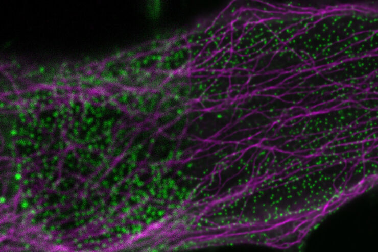
人工智能如何增强共聚焦成像
在本文中,我们将展示人工智能(AI)如何增强您的成像实验。即,由 Aivia 提供支持的动态信号增强如何在捕捉活细胞样本的时间动态的同时提高图像质量。
应用于显微术中的人工智能技术网络研讨会
我们展示了使用残差通道注意力网络(RCAN)还原和增强三维延时(4D)荧光显微数据。
细胞分析
先进的显微成像解决方案和用于细胞分析的人工智能分析软件有助于研究人员深入了解细胞功能和亚细胞结构。
