


 embedded in bulk vitreous ice (blue).")
 and tubulin (magenta), acquired using Viventis Deep. Courtesy of Akanksha Jain, Treutlein Lab ETH-DBSSE Basel (Switzerland).")

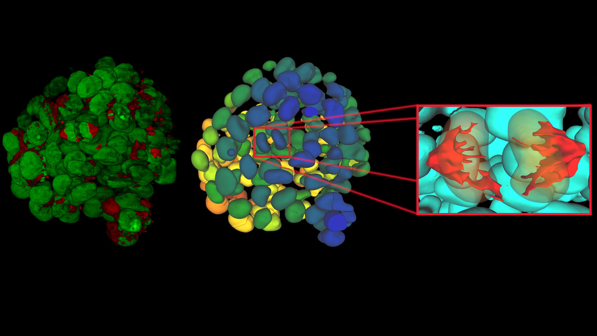
 2D slices of a 1 mm diameter midbrain neural organoid stained with DAPI (blue, nuclear stain), β-tubulin (green, neuronal stain), and GFAP (red, astrocyte stain).")


 and acceptor (A) molecule which participate in FRET (Förster resonance energy transfer).")

2026年2月25日至26日,徕卡显微系统公司首席执行官安妮特·林克博士作为德国经济代表团成员,随同德国联邦总理弗里德里希·梅尔茨对中国进行了国事访问。
阅读我们的最新文章
Cryo-ET Sample Preparation: From Waffle Method to Serial Lift-Out
Cryo-ET sample preparation becomes more demanding when specimens are thicker, larger, or more complex. This webinar brings together four perspectives on how high-pressure freezing can be connected…
如何深入了解类器官和细胞球模型
在本电子书中,您将了解3D细胞培养模型(如类器官和细胞球)成像的关键注意事项。探索创新型显微镜解决方案,来实时记录类器官和细胞球的动态成像过程。
Waffle Method Workflow: From HPF to Cryo-ET Lamellae
Waffle freezing provides an HPF-based route to cryo-ET sample preparation. This workflow guide follows the process from grid and carrier assembly to vitrification, cryo-FIB milling, lamella…
Fast, High-Contrast Widefield Imaging of Optically Challenging Samples
Live‑cell imaging of large, complex biological samples often requires large fields of view, sub-cellular resolution, high-sensitivity, and fast acquisition – all while maintaining low illumination…
Dental Loupes vs Microscopes: Exploring Visualization Options in Dentistry
Dental professionals often ask: “Should I use dental loupes or invest in a microscope?” This article explores the key differences between dental microscopes and dental loupes, focusing on…
Spatial Proteomics Workflow in Blood Cancer (MPNs)
Megakaryocytes play a central role in the biology of myeloproliferative neoplasms (MPNs), yet their in vivo proteomic characterization remains a major challenge due to low abundance and disrupted…
选择牙科显微镜时需考虑的六大特性
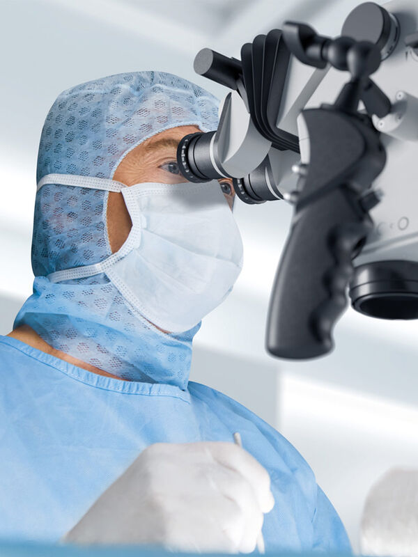
在牙医学中,手术显微镜对于进行高质量和成功的手术来说变得愈发重要,尤其是在牙髓病学领域。显微镜协助牙医进行微创手术,旨在保护牙质、保留组织、最大限度地降低风险并缩短愈合时间。
要选择适合牙医需求的显微镜,了解现代牙科显微镜的一些决定性特征将十分有帮助。
Multiscale Imaging of Organoids: High Content to Light Sheet
Learn multiscale organoid imaging: fixed high content phenotyping, gentle dual view light sheet, and reproducible pipelines that turn 3D data into insights.
比例成像
细胞的许多基本功能在很大程度上依赖于离子(例如钙、镁)、电压势和细胞质与周围细胞外空间之间的 pH 值的微妙但动态的平衡。这些平衡的变化会显著改变细胞的行为和功能。因此,实时测量细胞内离子、电压和 pH…
荧光寿命成像与荧光共振能量转移
荧光寿命是荧光团在发射荧光光子返回基态之前保持其激发态的平均时间长度。这取决于荧光团的分子组成和纳米环境。
FLIM将寿命测量与成像相结合:对每个图像像素以测得的荧光寿命进行颜色编码,产生额外的图像反差。因此,FLIM可以提供关于荧光分子空间分布的信息和有关其生化状态或纳米环境的信息。…

徕卡显微系统公司(Leica Microsystems)是丹纳赫旗下的一家公司,也是显微镜和科学仪器的领先供应商,该公司被全球公认的企业可持续发展评级机构 EcoVadis 授予银奖。

Leica Microsystems 宣布与 Fisher Scientific 建立新的战略和商业合作伙伴关系。
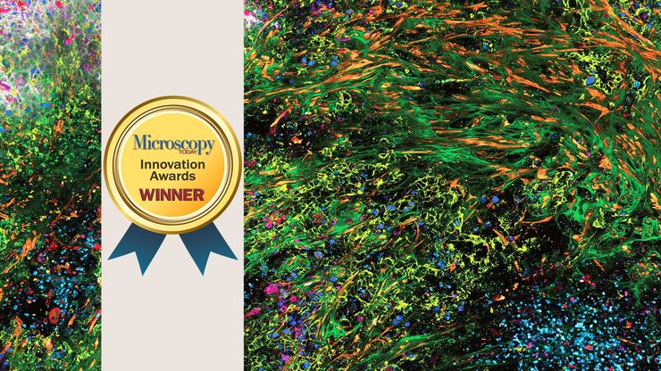
荣获《今日显微镜》创新奖的三维高倍成像复用解决方案能够深入揭示生物奥秘。

具有人工智能搜索功能的新平台可让客户方便地获得量身定制的显微镜解决方案
