


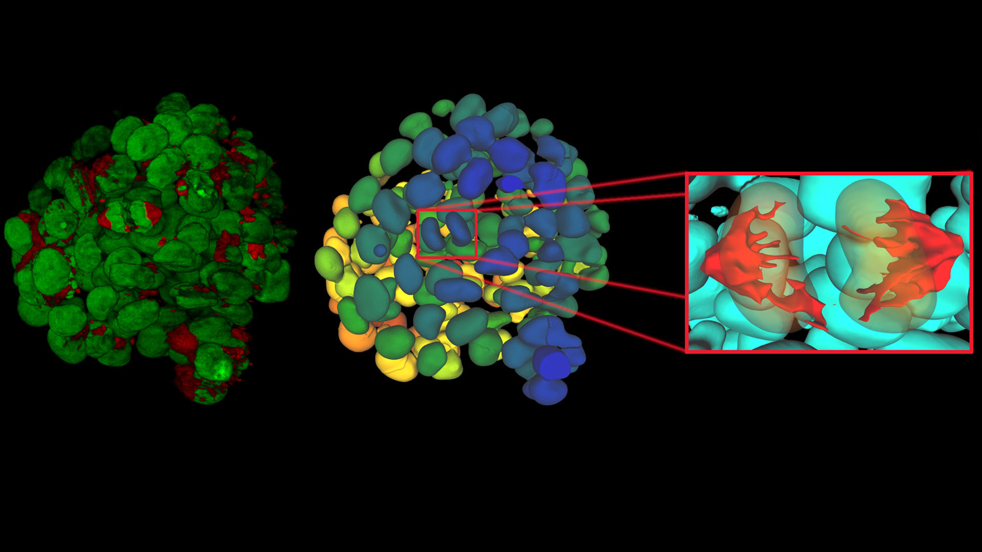


, insulin SGs (orange), microtubules (red), nucleus (yellow), and plasma membrane (transparent).")


, tubulin with Cy5 (red), and nuclei with DAPI (blue). Image courtesy of Dr. Günter Giese, Max Planck Institute for Medical Research, Heidelberg, Germany.")


2026年2月25日至26日,徕卡显微系统公司首席执行官安妮特·林克博士作为德国经济代表团成员,随同德国联邦总理弗里德里希·梅尔茨对中国进行了国事访问。
阅读我们的最新文章
4 Key Benefits of 3D Digital Microscopy in Ophthalmic Surgery
3D digital visualization is rapidly transforming ophthalmic surgery. Modern 3D surgical microscopes enable surgeons to perform procedures using high-resolution digital displays rather than traditional…
High-Pressure Freezing Protocols for Ultrastructural 3D EM
High pressure freezing (HPF) can help preserve hydrated cells and tissues close to their biological state at the moment of immobilization, supporting more reliable ultrastructural interpretation than…
Ultramicrotome UC Enuity in Practice: Stable 15 nm Sections at ZFE
After using the UCT and UC6 ultramicrotomes, Claudia Mayrhofer calls UC Enuity a leap in stability—so robust that vibrations and temperature shifts don’t spoil sections, even with multiple users. Auto…
利用偏光显微镜优势确保玻璃质量
玻璃是已知最古老的材料之一。如今,玻璃被广泛应用于各种领域,如光学仪器、门窗、太阳能电池板、食品、饮料和药品容器等,因此必须符合严格的玻璃质量标准,尤其是光学玻璃。利用偏光显微镜对平板玻璃、中空玻璃和压制玻璃进行质量控制既快捷又经济。无需进行耗时的样品制备,即可对结节、金属、晶体夹杂物和气泡等缺陷进行分析。
Expert Techniques for Superior Visualization in Cataract Surgery
Join renowned ophthalmic surgeons, Dr. Hussein Almuhtaseb and Mr. Simon Madge, as they share their clinical expertise and real-world surgical strategies during the 2025 Online Cataract Surgery…
消除激光显微切割中的静电干扰
激光显微切割(LMD)中的静电荷会导致两种严重问题:样品粘附在带电表面而丢失,或者样品飞入相邻的孔中造成交叉污染。我们在 Leica LMD7 环境室中集成了一个离子发生器除静电。离子发生器将静电位移从 16%(16/100 次切割)降低到 0.2%(1/450+ 次切割)。低吸附 384 孔板的收集率从 65-75% 提高到 85-95%。
History, Developments and Trends of Microscopy in Cancer Research
Cancer is a global disease, with 18 million new cases diagnosed and 10 million cancer-related deaths worldwide in 2020. This burden is set to increase, with a projected increase in cases of ~55% by…
荧光染料应用和特性概述
本文将介绍常用的荧光染料并概述其特性。荧光显微镜借助荧光染料、荧光蛋白或使用抗体的免疫荧光染色来研究特定的细胞成分。由于荧光剂种类繁多,荧光显微镜可用于检查蛋白质、核酸、聚糖、细胞器和其他细胞结构。
Researchers Insights: Microscopy in Cancer Research
Discover how imaging techniques are driving cancer research forward. In this issue, we present comprehensive multimodal studies using microscopy, as well as new directions in intraoperative cancer…
Predictive Service Prevents Downtime in Ghent
At the VIB BioImaging Core in Ghent, Belgium, researchers depend on Leica’s Stellaris 8 confocal microscope to explore the frontiers of biomedical science. When Leica’s RemoteCare system detected a…

徕卡显微系统公司(Leica Microsystems)是丹纳赫旗下的一家公司,也是显微镜和科学仪器的领先供应商,该公司被全球公认的企业可持续发展评级机构 EcoVadis 授予银奖。

Leica Microsystems 宣布与 Fisher Scientific 建立新的战略和商业合作伙伴关系。
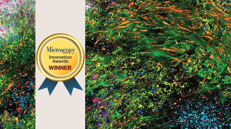
荣获《今日显微镜》创新奖的三维高倍成像复用解决方案能够深入揭示生物奥秘。

具有人工智能搜索功能的新平台可让客户方便地获得量身定制的显微镜解决方案
