Filter articles
标签
产品
Loading...
 labeled with membrane-permeable calcein, high-pressure frozen in salt water using EM ICE.")
High-Pressure Freezing for Organoids: Cryo CLEM & FIB Lift Out
Master cryo EM workflow steps for challenging 3D samples: when to choose HPF vs. plunge freezing, reproducible blotting/ice control, contamination aware transfers, Cryo CLEM 3D targeting in organoids,…
Loading...

用于三维生物成像的集成连续切片与冷冻电镜工作流程
本场网络研讨会探讨了集成化工具如何支持从样品制备到图像分析的电子显微镜全流程。专家Andreia Pinto博士、Adrian Boey博士与Hoyin Lai博士将介绍UC Enuity超薄切片机和Aivia图像分析平台,并演示这些工具如何同时适用于常温与低温实验环境。会议内容包含阵列断层成像、基于深度学习的图像分割、以及生物成像中cryo-lift-out工作流程的实际案例解析。
Loading...

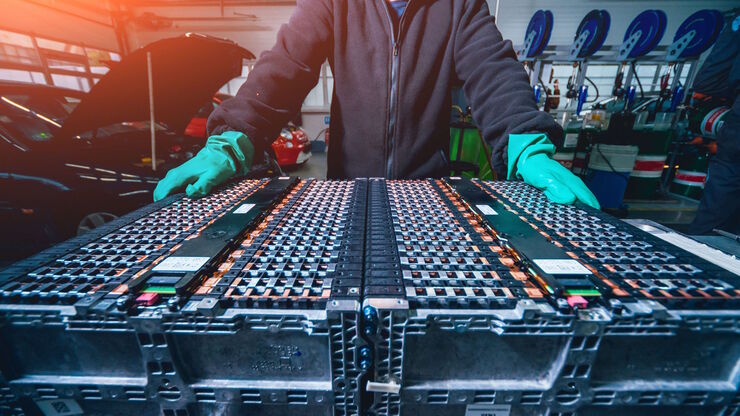
通过Cryo-EM(冷冻电镜)和 CryoFIB(冷冻聚焦离子束) 揭示钠电池退化机制
探索低温电镜和聚焦离子束技术如何揭示钠电池界面的内在结构。本次研讨会将提出基于隔膜渗透(而非枝晶生长)的新型退化模型,并解析电解液溶剂如何影响界面稳定性与电池性能。
Loading...

从显微镜到电镜:完整的冷冻光电联用工作流程
在题为“多模态玻璃化征程,从实验台到电子显微镜的冷冻关联工作流程”的网络研讨会上,专家团队(Edoardo D'Imprima、Zhengyi Yang、Andreia Pinto 和 Martin…
Loading...

通过自动切片改善您的超薄切片工作流程
在不断发展的电镜样品制备领域,保持领先地位至关重要。这个网络研讨会提供了关于超薄切片最新进展的重要见解,这些进展可以显著增强您实验室的能力。