

Image Gallery: THUNDER Imager
To help you answer important scientific questions, THUNDER Imagers eliminate the out-of-focus blur that clouds the view of thick samples when using camera-based fluorescence microscopes. They achieve…

激光显微切割耗材
激光显微切割(LMD)系统拥有多种不同类型的耗材,覆盖从基础到高度专业化的广泛应用领域,使科研人员能够根据自身研究选择个性化配置方案

FLIM( 荧光寿命成像)显微镜如何帮助检测微塑料污染
在生物样本中使用自发荧光是一种广泛应用的方法,可以帮助深入了解系统或生物体。这种特性不仅存在于生物系统中,人工材料如塑料也能够发出自发荧光。通过荧光寿命成像显微镜(FLIM)测量这种自发荧光的时间分辨率,可以获得荧光衰减的数据,即荧光寿命(τ)。我们的研究表明,荧光寿命可以用于无标记地表征塑料(微塑料)。
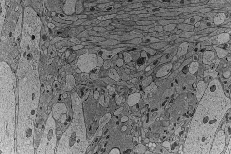
高压冷冻技术:揭示突触传递的功能机制
深入了解如何在 EM ICE 中应用光遗传学刺激技术,以及该技术如何有望揭示突触传递的结构与功能机制。获取关于如何将光遗传学刺激应用于小鼠急性脑切片和器官型脑切片培养物中完整神经网络的详细介绍。

冷冻电子显微镜的工作流程与仪器配置
冷冻电子显微镜作为一种日益流行的研究大分子复合体结构的模态,已在细胞生物学领域促成了众多新见解。近年来,该技术进一步向原位结构生物学方向拓展,成为解析自然状态下结构的首选技术。同样,冷冻断裂与冷冻扫描电子显微镜(SEM)的应用也日益广泛。

模式生物研究
模式生物是研究人员用来研究特定生物学过程的物种。 它们具有与人类相似的遗传特征,通常用于遗传学、发育生物学和神经科学等研究领域。 选择模式生物的原因通常是它们在实验室环境中易于保持和繁殖、生成周期短,或能够产生突变体来研究某些性状或疾病。
:内皮细胞,IsoB4(红色):血管,小胶质细胞抗 GFAP 抗体(蓝色):星形胶质细胞。样本由美国南旧金山 Genentech 公司的 Jeremy Burton")
消翳现真—突破传统宽场成像的极限
许多软件包都包含成像优化算法,通过降低背景噪声来增强图像特征的对比度。从 WF 图像中去除背景噪声最常用的方法是滚动球和滑动抛物面。近期徕卡显微系统公司推出了其自主研发的成像优化技术—即时成像解析(ICC),该技术已集成于所有徕卡THUNDER宽场成像平台。
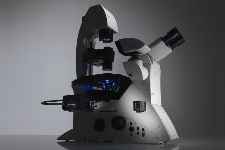
Factors to Consider When Selecting a Research Microscope
An optical microscope is often one of the central devices in a life-science research lab. It can be used for various applications which shed light on many scientific questions. Thereby the…
 - THUNDER Imager 3D Cell Culture Influenca virus – red, cilia – green, Nuclei – blue.")
免疫荧光如何帮助病毒学研究?
由于全球 COVID-19 大流行,现代病毒学研究变得比以往任何时候都更加重要。病毒学家可以应用许多强大的技术和检测方法来研究病毒的结构和功能。
