

Eliminating Electrostatic Interference in Laser Microdissection
Electrostatic charge in laser microdissection (LMD) causes two critical failures: samples stick to charged surfaces and are lost, or samples fly into adjacent wells and cause cross-contamination. We…

History, Developments and Trends of Microscopy in Cancer Research
Cancer is a global disease, with 18 million new cases diagnosed and 10 million cancer-related deaths worldwide in 2020. This burden is set to increase, with a projected increase in cases of ~55% by…
, tubulin with Cy5 (red), and nuclei with DAPI (blue). Image courtesy of Dr. Günter Giese, Max Planck Institute for Medical Research, Heidelberg, Germany.")
荧光染料
荧光显微镜的基本原理是借助荧光染料对细胞成分进行高度特异性的可视化观察。这可能是一种与兴趣蛋白质遗传相关的荧光蛋白,如绿色荧光蛋白(GFP)等。如果克隆无法实现,例如在组织学样本上无法实现,则需要使用另一种技术如免疫荧光染色来对兴趣蛋白质进行可视化观察。为此,人们使用抗体来连接不同的荧光染料并将其直接或间接地结合到适当的靶点上。此外,借助荧光染料,荧光显微镜的应用就不再仅局限于蛋白质观察,还能对核…
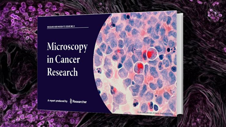
Researchers Insights: Microscopy in Cancer Research
Discover how imaging techniques are driving cancer research forward. In this issue, we present comprehensive multimodal studies using microscopy, as well as new directions in intraoperative cancer…
, Tropomyosin (cardiomyocytes, red) and GFP (primordial cardiac layer, green).")
显微镜中的荧光
荧光显微技术是一种特殊的光学显微镜技术。它利用的是荧光色素在一定波长的光激发下发光的能力。通过抗体染色或荧光蛋白标记,可以用这种荧光色素标记感兴趣的蛋白质。这样就可以确定单分子物种的分布、数量及其在细胞内的定位。此外,还可以进行共定位和相互作用研究,使用可逆结合染料(如 Ca2+ 和 fura-2)观察离子浓度,以及观察细胞的内吞和外吞过程。如今,利用荧光显微镜甚至可以对亚分辨率颗粒进行成像。
 labeled with membrane-permeable calcein, high-pressure frozen in salt water using EM ICE.")
High-Pressure Freezing for Organoids: Cryo CLEM & FIB Lift Out
Master cryo EM workflow steps for challenging 3D samples: when to choose HPF vs. plunge freezing, reproducible blotting/ice control, contamination aware transfers, Cryo CLEM 3D targeting in organoids,…
 and astrocytes (green) in a cortical spheroid derived from human induced pluripotent stem cells.")
活细胞成像指南
在生命科学各研究领域的广泛应用中,活细胞成像是一种不可或缺的工具,用于观察细胞在尽可能接近活体(即活的、活跃的)状态下的情况。本指南回顾了确保成功进行活细胞成像的各种重要注意事项,并介绍了各种旨在克服常见挑战的高性能解决方案。这些进展使我们能够对细胞生理学和动力学有新的认识。
选择研究用显微镜时应考虑的因素
光学显微镜通常是生命科学研究实验室的核心设备之一。它可用于各种应用,揭示许多科学问题。因此,显微镜的配置和功能对其应用范围至关重要,从明视野显微镜到荧光显微镜,再到活细胞成像。本文简要概述了显微镜的相关功能,并总结了在选择研究用显微镜时应考虑的关键问题。
 image of a cross section of C. elegans (roundworm). Courtesy of T. Müller-Reichert, MPI-CBG, Dresden, Germany and K. McDonald, University of California, Berkeley, USA.")
高压冷冻简介
水是细胞最主要的组成部分,因此对于维持细胞超微结构至关重要。目前,冷冻固定是固定细胞成分,而不导致其显著结构变化的唯一途径。现阶段有两种常见的方法:投入冷冻与高压冷冻固定。
