Filter articles
标签
产品
Loading...

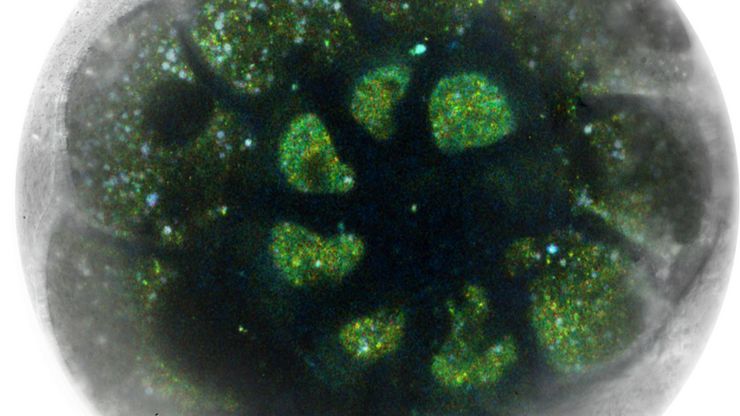
High-Pressure Freezing for Organoids: Cryo CLEM & FIB Lift Out
Master cryo EM workflow steps for challenging 3D samples: when to choose HPF vs. plunge freezing, reproducible blotting/ice control, contamination aware transfers, Cryo CLEM 3D targeting in organoids,…
Loading...

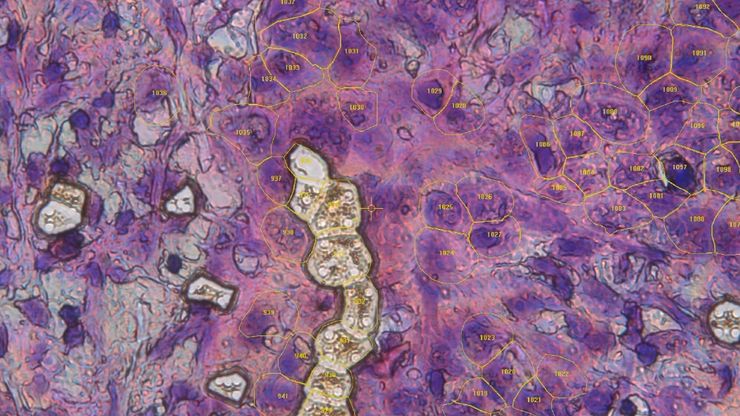
人工智能与深度视觉蛋白质组学 (DVP) 相结合,推进疾病研究
在这次网络研讨会上,Andreas Mund 博士将介绍深度可视蛋白质组学(DVP)--一种将人工智能驱动的组织空间分辨、非靶向蛋白质组学相结合的尖端平台。他展示了 DVP 如何从最小的、表型匹配的细胞群中识别数千种蛋白质,并在复杂的临床组织样本中生成高分辨率分子图谱,从而在细胞水平上解码疾病机制。
Loading...

罕见疾病 CRISPR 疗法的开发与风险解除
Fyodor Urnov博士和Sadik Kassim博士最初是在ASGCT 2025会议上作这一按需演讲的,演讲的重点是遗传医学中的一个关键挑战:如何将CRISPR疗法从单一疾病解决方案扩展到平台方法,特别是针对罕见的儿科遗传疾病。Urnov 博士展示了由 Matthew Kan 博士领导的创新基因组研究所的工作,这是 IGI-Danaher Beacon for CRISPR Cures…
Loading...

用于三维生物成像的集成连续切片与冷冻电镜工作流程
本场网络研讨会探讨了集成化工具如何支持从样品制备到图像分析的电子显微镜全流程。专家Andreia Pinto博士、Adrian Boey博士与Hoyin Lai博士将介绍UC Enuity超薄切片机和Aivia图像分析平台,并演示这些工具如何同时适用于常温与低温实验环境。会议内容包含阵列断层成像、基于深度学习的图像分割、以及生物成像中cryo-lift-out工作流程的实际案例解析。
Loading...

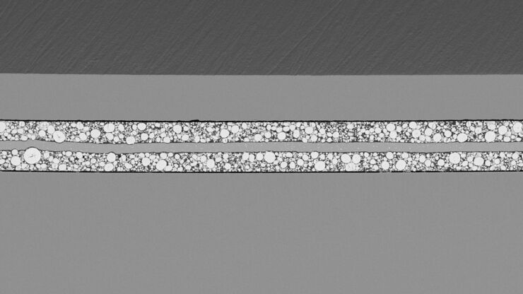
通过Cryo-EM(冷冻电镜)和 CryoFIB(冷冻聚焦离子束) 揭示钠电池退化机制
探索低温电镜和聚焦离子束技术如何揭示钠电池界面的内在结构。本次研讨会将提出基于隔膜渗透(而非枝晶生长)的新型退化模型,并解析电解液溶剂如何影响界面稳定性与电池性能。
Loading...

如何优化多标成像技术推动3D空间组学发展
本次网络研讨会上,徕卡显微系统的Julia Roberti博士与Luis Alvarez博士将介绍STELLARIS共聚焦平台的全新功能SpectraPlex,该技术可实现超多标三维空间成像。该技术旨在通过实现超多标成像且无需频繁人工干预,从而简化和增强空间生物学应用。
