生命科学研究

生命科学研究

生命科学研究

在生命科学研究中心,您可以掌握最新的关于先进显微镜、成像技术、电镜样品制备和图像分析的前沿应用和创新,涵盖的主题包括细胞生物学、神经科学和癌症研究。希望在这里可以帮助您提升研究能力和精进显微镜在各个科学领域实际应用,并了解徕卡如何通过精确的可视化、图像解读和推进研究进展来赋能您的工作。

Integrated Serial Sectioning and Cryo-EM Workflows for 3D Biological Imaging

This on-demand webinar explores how integrated tools can support electron microscopy workflows from sample preparation to image analysis. Experts Andreia Pinto, Adrian Boey, and Hoyin Lai present the…
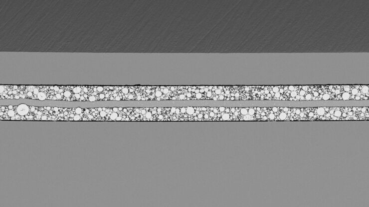
SEM image of the full Li-NMC electrode sample, showing the two porous layers and the metal film at the center of the structure.

Revealing Sodium Battery Degradation via Cryo-EM and CryoFIB

Explore how cryogenic electron microscopy and focused ion beam techniques uncover the intrinsic structure of sodium battery interfaces. This webinar presents a new degradation model based on separator…
Mouse hippocampus brain slice on a grid after HPF using the “Waffle Method”.

The “Waffle Method”: High-Pressure Freeze Complex Samples

This article describes the advantages of a special high pressure freezing method, the so-called “Waffle Method”. Learn how the “Waffle Method” uses EM grids as spacers for high-pressure freezing,…
UC Enuity with Diatome diamond knife.

Mastering Polymer Sectioning with Helmut Gnaegi

When it comes to ultramicrotomy, few names carry the weight of Helmut Gnaegi. As co-founder of Diatome, a global leader in diamond knife technology, Helmut has spent decades refining the art and…
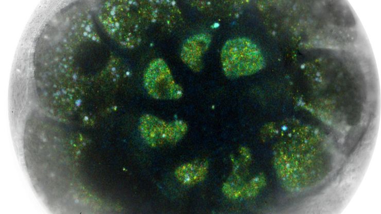
C. elegans embedded in Lowicryl® HM20; pharynx showing red fluorescence (mCherry). The overview shows a front view onto the resin capsule formed by the bottom of a flow-through chamber of the EM AFS2. The capsule was pretrimmed manually. The blockface was trimmed automatically using the AutoTrim function of UC Enuity guided by fluorescence of the worm. Edge length of both squares in relation to the images is 250 µm.

How Fluorescence Guides Sectioning of Resin-embedded EM Samples

Electron microscopes, including transmission electron microscopes (TEM) and scanning electron microscopes (SEM), are widely utilized to gain detailed structural information about biological samples or…
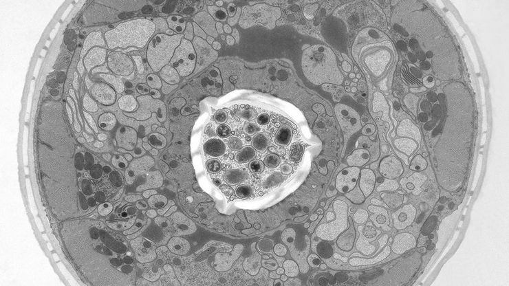
嵌入在Epon环氧树脂中的秀丽隐杆线虫,与四氧化锇对比。树脂块经手工预修整。

如何通过自动化超薄切片技术节省时间与样本

本文阐述了如何利用树脂包埋电镜样本的 3D micro-CT 数据,在切片前将样本修整至预设目标平面。采用Leica UC Enuity 系统的交互式自动化方案,可显著节省时间、减少样本损耗及缩短新手用户的培训周期。
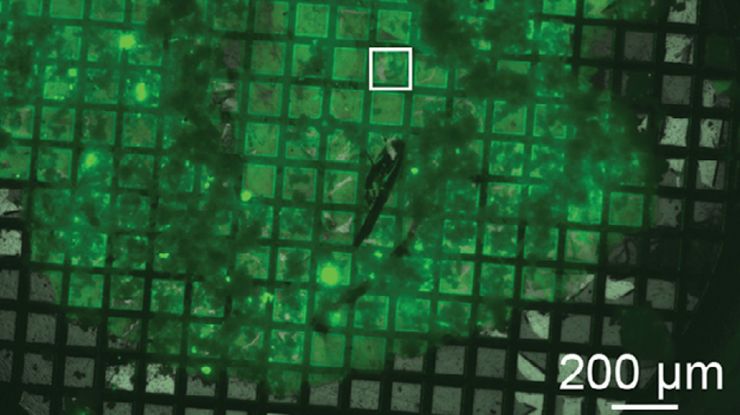
Complete camera overview of EM grid recorded with 3 channels. Inserts displaying the positions, where superresolved 3D confocal images were recorded. 3D renderings of these positions are shown in the zoomed inserts. Fluorescence channels (nuclei by Hoechst, blue; mitochondria by MitoTracker Green, green; lipid Droplets by Bodipy and Crimson Beads, red). Width of a grid square is 90 ?m, width of a grid bar is 35 ?m. Samples kindly provided by Ievgeniia Zagoriy, Mahamid-Group, EMBL Heidelberg, Germany.

从显微镜到电镜:完整的冷冻光电联用工作流程

在题为“多模态玻璃化征程,从实验台到电子显微镜的冷冻关联工作流程”的网络研讨会上,专家团队(Edoardo D'Imprima、Zhengyi Yang、Andreia Pinto 和 Martin…
Block-face created by automatic trimming under fluorescence. Mammalian cells of interest, stained with CellTrackerTM Green are visualized within the block-face using the UC Enuity equipped with the stereo microscope M205 FA. In the background a carbon finder grid in black is visible. All samples in the article are created by Felix Gaedke, PhD, CECAD, Cologne, Germany.

如何在块面中自动获取感兴趣的荧光细胞

本文介绍了使用超薄切片超薄切片机自动修整修块功能,获取树脂块面中带有荧光信号的细胞结构。我们展示了如何使用配置有体视显微镜 M205 FA 的超薄切片超薄切片机 UC Enuity ,来识别感兴趣的荧光细胞,如何自动修整包含细胞的块面,以及如何在切片中观察细胞而无需转移到外部显微镜。
UC Enuity

通过自动切片改善您的超薄切片工作流程

在不断发展的电镜样品制备领域,保持领先地位至关重要。这个网络研讨会提供了关于超薄切片最新进展的重要见解,这些进展可以显著增强您实验室的能力。
Scroll to top