生命科学研究

生命科学研究

生命科学研究

在生命科学研究中心,您可以掌握最新的关于先进显微镜、成像技术、电镜样品制备和图像分析的前沿应用和创新,涵盖的主题包括细胞生物学、神经科学和癌症研究。希望在这里可以帮助您提升研究能力和精进显微镜在各个科学领域实际应用,并了解徕卡如何通过精确的可视化、图像解读和推进研究进展来赋能您的工作。
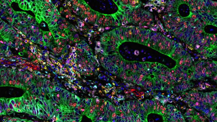
Cell DIVE multiplexed image of FFPE tissue section from human colon adenocarcinoma tissue.

Multiplexed Imaging Reveals Tumor Immune Landscape in Colon Cancer

Cancer immunotherapy benefits few due to resistance and relapse, and combinatorial therapeutic strategies that target multiple steps of the cancer-immunity cycle may improve outcomes. This study shows…
Cell DIVE multiplexed image of FFPE tissue section from human invasive ductal carcinoma (IDC)

AI-Powered Hi-Plex Spatial Analysis Tools for Breast Cancer Research

Breast cancer (BC) is the leading cause of cancer-related deaths in women. Investigating the tumor microenvironment (TME) is crucial to elucidate the mechanisms of tumor progression. Systematic…
Automated Laser Microdissection for Proteome Analysis

深度视觉蛋白质组学提供精确的空间蛋白质组信息

尽管可使用基于成像和质谱的方法进行空间蛋白质组学研究,但是图像与单细胞分辨率蛋白丰度测量值的关联仍然是个巨大的挑战。最近引入的一种方法,深层视觉蛋白质组学(DVP),将细胞表型的人工智能图像分析与自动化的单细胞或单核激光显微切割及超高灵敏度的质谱分析结合在了一起。DVP在保留空间背景的同时,将蛋白丰度与复杂的细胞或亚细胞表型关联在一起。
Multicolor TauSTED Xtend 775 for Cell Biology applications that require nanoscopy resolution for multiple cellular components. Cells showing vimentin fibrils (AF 594), actin network (ATTO 647N), and nuclear pore basket (CF 680R). Sample courtesy of Brigitte Bergner, Mariano Gonzales Pisfil, Steffen Dietzel, Core Facility Bioimaging, Biomedical Center, Ludwig-Maximilians-University, Munich, Germany.

STED样品制备指南

这份指南旨在帮助用户优化受激发射损耗(STED)纳米成像的样品制备,特别是在使用徕卡微系统的STED显微镜时。它提供了单色STED成像用荧光标记的概述,并对其性能进行了评级。
Image of immunofluorescently labelled cells where mitochondria are indicated with red, nuclei with blue, and actin with green.

采用徕卡THUNDER-DM6B观察SARS-CoV-2感染宿主细胞及其复制过程

冠状病毒2致重度急性呼吸综合征(SARS-CoV-2) 冠状病毒2致重度急性呼吸综合征(SARS-CoV-2)出现于2019年末,并快速传播全世界。由于其大面积的影响,研究人员对病毒的性质进行了深入的研究以期最终阻止大流行。一个重要的方面是病毒如何在宿主细胞中复制。Ogando及其同事的研究已经揭示了SARS-CoV-2的复制动力学、适应能力和细胞病理学。他们的工具之一是用荧光显微镜观察SARS…
Cell counts for each biomarker were divided by total number of cells to give a percentage of biomarker positive cells out of total cells for each biomarker.

改善空间生物学研究可重复性的方法

利用自动化、高质量抗体以及经验证的多重成像工作流程,Cell DIVE能够提供可重复的实验结果。
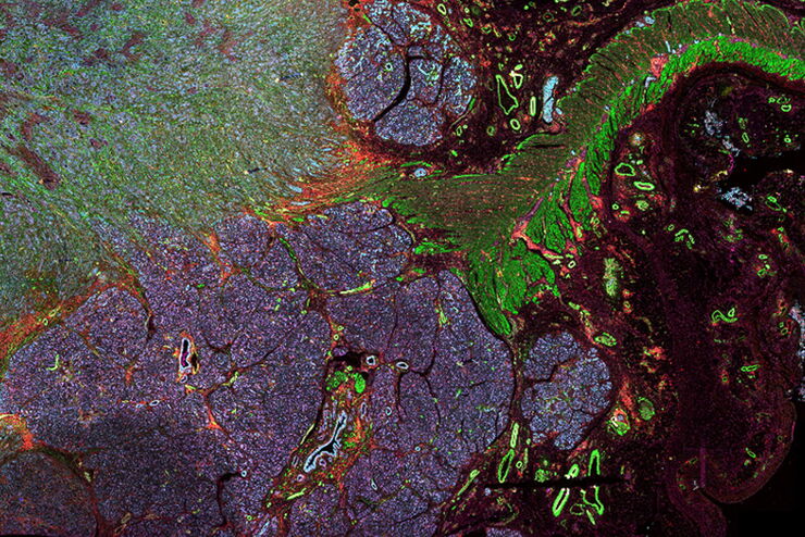
PDAC Multiplexed imaging of CST panels enables an examination of immune cell components in pancreatic ductal adenocarcinoma (IPDAC) tissue on a single slide.

表征肿瘤环境以揭示洞察和空间分辨率

肿瘤环境的表征可以为癌症进展和潜在治疗靶点提供更深入的见解。我们已经使用来自Cell Signaling Technology(CST)的各种IHC验证抗体,在胰腺癌的Cell DIVE研究中验证了30多种偶联抗体。
Pancreatic Ductal Adenocarcinoma with 5 biomarkers shown – SMA, PanCK PCK26, PanCK AE1, Vimentin, and Glut1.

借助多重成像深入了解胰腺癌的复杂性

胰腺癌是一种很难治疗的肿瘤疾病。Cell DIVE多重成像可以视觉呈现30种生物标志物以探测胰管癌的微环境。此面板可以检查肿瘤组织多个层级的问题,包括淋巴细胞、血管新生、转移、侵袭、炎症、缺氧、代谢和免疫。多重成像和分析可以对肿瘤组织中的许多生物活动提供更为深入的洞察信息,从而可以深入研究这些信息。
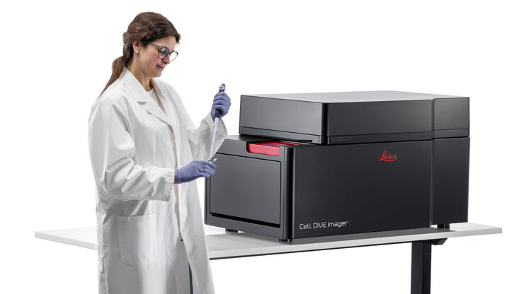
Cell DIVE Multiplex Imaging Solution

化繁为简:多重成像中的抗体

了解抗体对于多重成像研究的重要意义,以及如何规划并建立自己的抗体组合
Scroll to top