
Filter articles
标签
Loading...

生理学图片库
生理学是关于生物体内的过程和功能。生理学研究的重点是生物体器官、组织或细胞的活动和功能,包括所涉及的物理和化学现象。在此,我们以不同的样本为例,向您展示与生理学有关的图片。
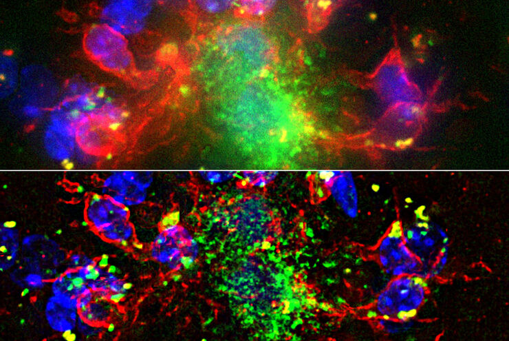
 and astrocytes (green) in a cortical spheroid derived from human induced pluripotent stem cells.")

Loading...

自适应反卷积与 Computational Clearing 结合的力量
反卷积是一种计算方法,用于恢复被点扩散函数(PSF)和噪声源破坏的物体图像。在本技术简介中,您将了解徕卡显微系统提供的反卷积算法如何帮助您克服宽视场 (WF) 荧光显微镜中由于光的波动性和光学元件对光的衍射而造成的图像分辨率和对比度损失。探索由用户控制或自动反卷积的方法,查看并解析更多的结构细节。
Loading...

Image Gallery: THUNDER Imager
To help you answer important scientific questions, THUNDER Imagers eliminate the out-of-focus blur that clouds the view of thick samples when using camera-based fluorescence microscopes. They achieve…
Loading...
:内皮细胞,IsoB4(红色):血管,小胶质细胞抗 GFAP 抗体(蓝色):星形胶质细胞。样本由美国南旧金山 Genentech 公司的 Jeremy Burton")
消翳现真—突破传统宽场成像的极限
许多软件包都包含成像优化算法,通过降低背景噪声来增强图像特征的对比度。从 WF 图像中去除背景噪声最常用的方法是滚动球和滑动抛物面。近期徕卡显微系统公司推出了其自主研发的成像优化技术—即时成像解析(ICC),该技术已集成于所有徕卡THUNDER宽场成像平台。
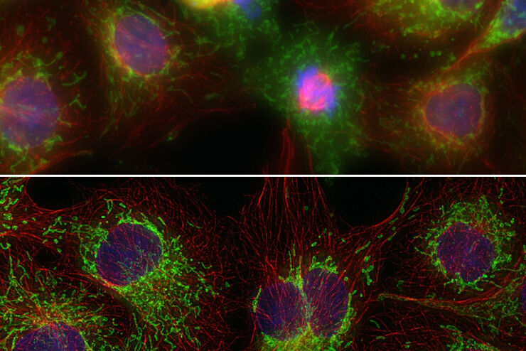
 - THUNDER Imager 3D Cell Culture Influenca virus – red, cilia – green, Nuclei – blue.")
Loading...
Computational Clearing - 增强3D标本成像
本次网络研讨会旨在阐明有助于THUNDER显微成像仪实现三维样品可视化的关键规格,并改进研究人员的成像相关工作流程。
Loading...
THUNDER成像:高效、灵活、易操作,让您的日常成像工作流更轻松
本次网络研讨会将展示 THUNDER 在许多不同生命科学应用中的多功能性和性能:从计数视网膜切片中的细胞核和癌组织切片中的 RNA 分子,到监测阿拉伯芥幼苗中的钙波等等。
