需要帮助
如你需要正置显微镜解决方案,请联系我们。
related articles
 2D slices of a 1 mm diameter midbrain neural organoid stained with DAPI (blue, nuclear stain), β-tubulin (green, neuronal stain), and GFAP (red, astrocyte stain).")
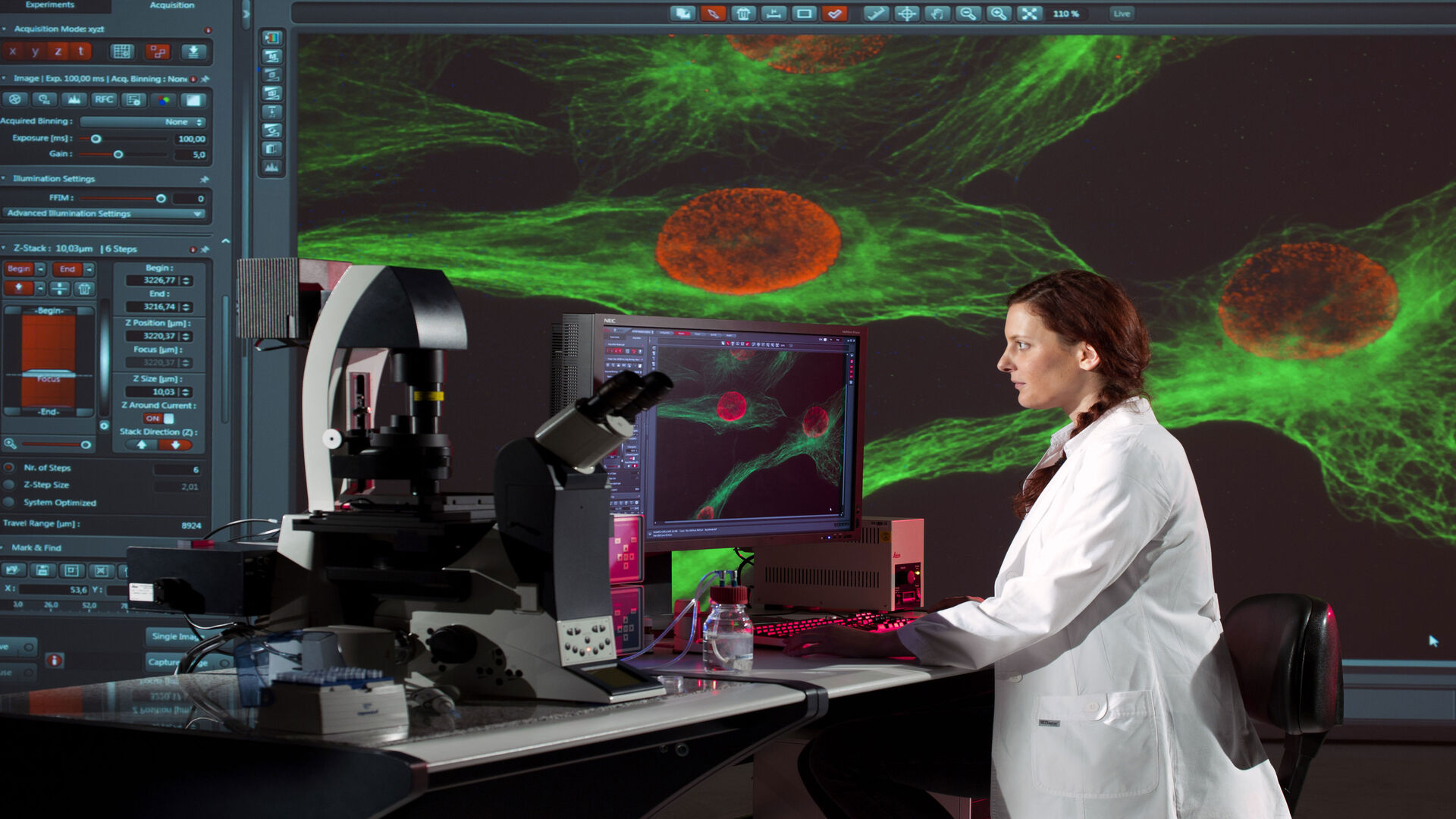
Fast, High-Contrast Widefield Imaging of Optically Challenging Samples
Live‑cell imaging of large, complex biological samples often requires large fields of view, sub-cellular resolution, high-sensitivity, and fast acquisition – all while maintaining low illumination…

Dental Loupes vs Microscopes: Exploring Visualization Options in Dentistry
Dental professionals often ask: “Should I use dental loupes or invest in a microscope?” This article explores the key differences between dental microscopes and dental loupes, focusing on…

Spatial Proteomics Workflow in Blood Cancer (MPNs)
Megakaryocytes play a central role in the biology of myeloproliferative neoplasms (MPNs), yet their in vivo proteomic characterization remains a major challenge due to low abundance and disrupted…

选择牙科显微镜时需考虑的六大特性
在牙医学中,手术显微镜对于进行高质量和成功的手术来说变得愈发重要,尤其是在牙髓病学领域。显微镜协助牙医进行微创手术,旨在保护牙质、保留组织、最大限度地降低风险并缩短愈合时间。
要选择适合牙医需求的显微镜,了解现代牙科显微镜的一些决定性特征将十分有帮助。

Multiscale Imaging of Organoids: High Content to Light Sheet
Learn multiscale organoid imaging: fixed high content phenotyping, gentle dual view light sheet, and reproducible pipelines that turn 3D data into insights.

比例成像
细胞的许多基本功能在很大程度上依赖于离子(例如钙、镁)、电压势和细胞质与周围细胞外空间之间的 pH 值的微妙但动态的平衡。这些平衡的变化会显著改变细胞的行为和功能。因此,实时测量细胞内离子、电压和 pH…
 and acceptor (A) molecule which participate in FRET (Förster resonance energy transfer).")
荧光寿命成像与荧光共振能量转移
荧光寿命是荧光团在发射荧光光子返回基态之前保持其激发态的平均时间长度。这取决于荧光团的分子组成和纳米环境。
FLIM将寿命测量与成像相结合:对每个图像像素以测得的荧光寿命进行颜色编码,产生额外的图像反差。因此,FLIM可以提供关于荧光分子空间分布的信息和有关其生化状态或纳米环境的信息。…

4 Key Benefits of 3D Digital Microscopy in Ophthalmic Surgery
3D digital visualization is rapidly transforming ophthalmic surgery. Modern 3D surgical microscopes enable surgeons to perform procedures using high-resolution digital displays rather than traditional…
, insulin SGs (orange), microtubules (red), nucleus (yellow), and plasma membrane (transparent).")
High-Pressure Freezing Protocols for Ultrastructural 3D EM
High pressure freezing (HPF) can help preserve hydrated cells and tissues close to their biological state at the moment of immobilization, supporting more reliable ultrastructural interpretation than…
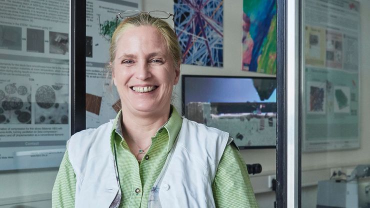
Ultramicrotome UC Enuity in Practice: Stable 15 nm Sections at ZFE
After using the UCT and UC6 ultramicrotomes, Claudia Mayrhofer calls UC Enuity a leap in stability—so robust that vibrations and temperature shifts don’t spoil sections, even with multiple users. Auto…