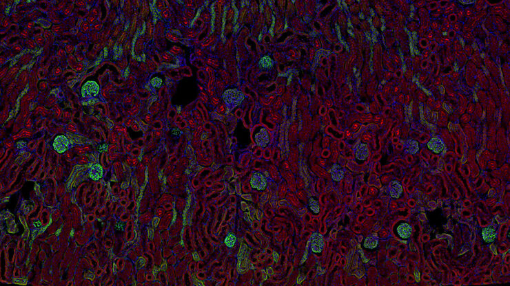
SpectraPlex 用于空间探索的3D多标成像解决方案

您的空间探索之旅从这里开始

阅读我们的最新文章

Virally labeled neurons (red) 
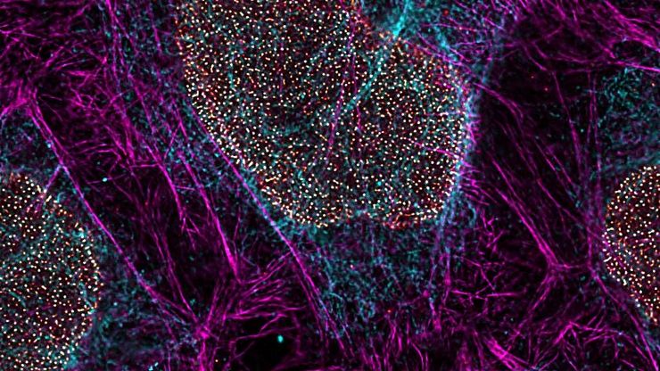
and astrocytes (green) in a cortical spheroid derived from human induced pluripotent stem cells. THUNDER Model Organism Imager with a 2x, 0.15 NA objective at 3.4x zoom was used to produce this 425 µm Z-stack (26 positions) which is presented here as an Extended Depth of Field (EDoF) projection. Images courtesy of Dr. F. Birey, Dr. S. Pasca laboratory, Palo Alto, CA.

活细胞成像指南

在生命科学各研究领域的广泛应用中,活细胞成像是一种不可或缺的工具,用于观察细胞在尽可能接近活体(即活的、活跃的)状态下的情况。本指南回顾了确保成功进行活细胞成像的各种重要注意事项,并介绍了各种旨在克服常见挑战的高性能解决方案。这些进展使我们能够对细胞生理学和动力学有新的认识。
用 GFAP-A647 进行免疫染色并使用THUNDER成像仪组织成像的小鼠脑片。美国费城宾夕法尼亚大学 H. Xu 提供。

神经科学研究指南

神经科学通常需要研究具有挑战性的标本,以更好地了解神经系统和疾病。徕卡显微镜帮助神经科学家深入了解神经元功能。
3D high-plex imaging in cancer immunology. Overview of a pancreatic tumor section in mouse model, labeled with 15 markers and imaged in one go using STELLARIS with SpectraPlex. (https://www.nature.com/articles/d42473-024-00260-7)

如何优化多标成像技术推动3D空间组学发展

本次网络研讨会上,徕卡显微系统的Julia Roberti博士与Luis Alvarez博士将介绍STELLARIS共聚焦平台的全新功能SpectraPlex,该技术可实现超多标三维空间成像。该技术旨在通过实现超多标成像且无需频繁人工干预,从而简化和增强空间生物学应用。
Pancreatic Ductal Adenocarcinoma with 11 Apoptosis biomarkers shown – BAK, BAX, BCL2, BCLXL, Caspase9, CIAP1, NaKATPase, PCK26, SMAC, Vimentin, and XIAP.

利用空间蛋白质组学工作流程改革研究工作

空间蛋白质组学是《自然-方法》2024 年度方法,正在推动癌症、免疫学等领域的研究进展。通过将定位数据与组织中蛋白质的高通量成像结合起来,研究人员可以发现疾病进展和治疗反应方面的洞察力,从而更好地了解人类生物学。在这里,您可以了解更多有关空间生物学的信息,以及徕卡显微系统的工具如何推动蛋白质生物标记的可视化和分析取得进展。
Pancreatic Ductal Adenocarcinoma imaged with Cell DIVE. Analysis done by Aivia.

空间生物学指南

什么是空间生物学?在后组学时代,研究人员如何利用空间生物学工具来满足生物学问题日益增长的需求?本文简要概述了空间生物学及其技术,以及这一快速发展中的领域的关键研究问题。

癌症研究

癌症是一种复杂的异质性疾病,由于细胞生长失控而引起。 一个或一组细胞的基因和表观遗传的变化破坏了正常功能,导致细胞自发、不受控制地生长和增殖。
3D high-plex imaging in Cancer Immunology. Detail of a pancreatic tumor section in mouse model, labeled with 15 markers and imaged in one go using STELLARIS SpectraPlex. Total imaged volume 3.2 mm x 2.2 mm x 70 mm in 1 h 20 min. Source: 3D high-multiplex imaging in cancer immunology. Kunz L., Speziale D., et al., Nat. Methods (2024).

Advanced Tissue Imaging & Analysis

Gain insights into tissue structure and function to improve your understanding of spatial biology and disease mechanisms with advanced imaging solutions from Leica Microsystems.
Super-resolution image of intracellular structures using a STELLARIS STED microscope and TauSTED Xtend. The magenta color shows actin filaments stained with ATTO 647N.

Cellular Analysis

Advanced microscopy imaging solutions and AI-powered analysis software for cellular analysis helps researchers gain deeper insight into cellular functions and subcellular structures.

应用领域

生命科学研究

徕卡(Leica)生命科学显微镜凭借先进的创新和专业技术能力,支持观察、测量和分析微结构的成像要求。徕卡显微系统对科学应用领域的高度关注,使徕卡显微系统的用户始终保持领先位置

生物制药

对于生物制药行业,Leica 解决方案有助于加快药物发现,增强细胞分析,并支持符合法规的数据完整性。
Scroll to top