生命科学研究

生命科学研究

生命科学研究

在生命科学研究中心,您可以掌握最新的关于先进显微镜、成像技术、电镜样品制备和图像分析的前沿应用和创新,涵盖的主题包括细胞生物学、神经科学和癌症研究。希望在这里可以帮助您提升研究能力和精进显微镜在各个科学领域实际应用,并了解徕卡如何通过精确的可视化、图像解读和推进研究进展来赋能您的工作。

A Novel Laser-Based Method for Studying Optic Nerve Regeneration

Optic nerve regeneration is a major challenge in neurobiology due to the limited self-repair capacity of the mammalian central nervous system (CNS) and the inconsistency of traditional injury models.…
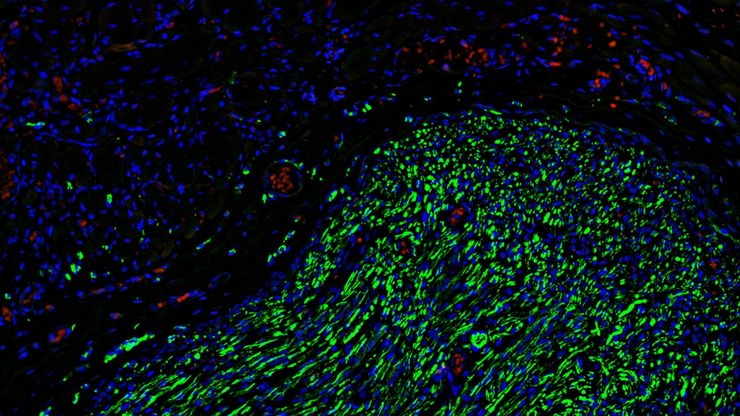
Fluorescence microscopy of sectioned tissue, showing the interface between the extensor digitorum longus muscle and the common peroneal nerve in the adult rat. Regenerative peripheral nerve interface (RPNI) at 2 weeks. Image acquired using Mica. Stained for nuclei (blue), neurofilaments (green) and S100B (red). Image courtesy of Dr. Aaron Lee, Department of Bioengineering (Lab of Dr. Rylie Green), Imperial College London.

How to Image Axon Regeneration in Deep Muscle Tissue

This study highlights Dr. Aaron Lee’s research on mapping nerve regeneration in muscle grafts post-amputation. Limb loss often leads to reduced quality of life, not only from tissue loss but also due…
A fruit fly (Drosophila melanogaster) observed with an Ivesta 3 stereo microscope during fly pushing (sorting of the flies). The scale bar length is 1 mm. Image courtesy of M. Benton, EMBL, Heidelberg, Germany.

A Guide to Using Microscopy for Drosophila (Fruit Fly) Research

The fruit fly, typically Drosophila melanogaster, has been used as a model organism for over a century. One reason is that many disease-related genes are shared between Drosophila and humans. It is…

用于三维生物成像的集成连续切片与冷冻电镜工作流程

本场网络研讨会探讨了集成化工具如何支持从样品制备到图像分析的电子显微镜全流程。专家Andreia Pinto博士、Adrian Boey博士与Hoyin Lai博士将介绍UC Enuity超薄切片机和Aivia图像分析平台,并演示这些工具如何同时适用于常温与低温实验环境。会议内容包含阵列断层成像、基于深度学习的图像分割、以及生物成像中cryo-lift-out工作流程的实际案例解析。
Blood vessel system of a zebrafish larvae

克服显微镜成像移动斑马鱼幼虫时的挑战

Zebrafish is a valuable model organism with many beneficial traits. However, imaging a full organism poses challenges as it is not stationary. Here, this case study shows how zebrafish larvae can be…
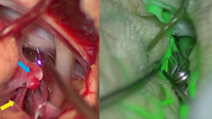
GLOW800 Augmented Reality Fluorescence used for real-time blood flow visualization in aneurysm clipping surgery

动脉瘤夹闭:使用 AR 荧光实时评估穿支血管

本文涵盖了两个动脉瘤夹闭案例,基于日本昭和大学医院神经外科主任水谷徹教授的见解,突显了 GLOW800 增强现实荧光在神经外科中的临床益处。它展示了神经外科医生如何在动脉瘤夹闭和其他复杂神经外科技术中,以自然色彩和深度感知的方式实时可视化与解剖结构相关的血流。
小鼠子宫内膜类器官的免疫荧光图像(CK14 和 DAPI 染色)

利用子宫内膜类器官推进子宫再生疗法

康教授团队致力于研究决定子宫微环境的关键因素,该环境对胚胎着床和妊娠维持至关重要。他们正为罹患阿什曼综合征等子宫内膜疾病的患者开发恢复子宫内膜功能的新型治疗策略。通过将 3D 子宫内膜类器官移植至小鼠模型,该团队揭示了子宫内膜强大的再生能力的细胞与分子机制。本次访谈将深入探讨其团队的研究内容及Mica在研究中所发挥的重要作用。
Optical microscope image, which is a composition of both brightfield and fluorescence illumination, showing organic contamination on a wafer surface. The inset images in the upper left corner show the brightfield image (above) and fluorescence image (below with dark background).

晶圆上的光刻胶残留和有机污染物的可视化

随着半导体上集成电路(IC)的尺寸低于10纳米,在晶圆检测中有效检测光刻胶残留等有机污染物和缺陷变得越来越重要。光学显微镜仍然是常见的检测方法,但对于有机污染物,明场和其他类型的照明可能会存在局限性。本文讨论了荧光显微镜如何在半导体行业的QC、故障分析和研发过程中有效检测晶圆上的光刻胶残留和其他有机污染物。

激光显微切割技术用于组织和细胞分离的协议 - 免费下载电子书

激光显微切割(LMD,也称为激光捕获显微切割或LCM)使用户能够分离特定的单个细胞或整个组织区域,甚至亚细胞结构如染色体。纯化的组织和细胞可用于下游的RNA、DNA和蛋白质组工作流程。
Scroll to top