AI meets Deep Visual Proteomics (DVP) to Advance Disease Research
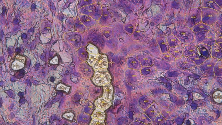
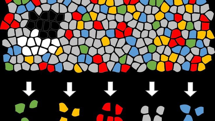
In this webinar, Dr. Andreas Mund introduces Deep Visual Proteomics (DVP) – a cutting-edge platform that integrates AI-powered tissue modeling with spatially resolved, untargeted proteomics. He…
Microscopy and AI Solutions for 2D Cell Culture
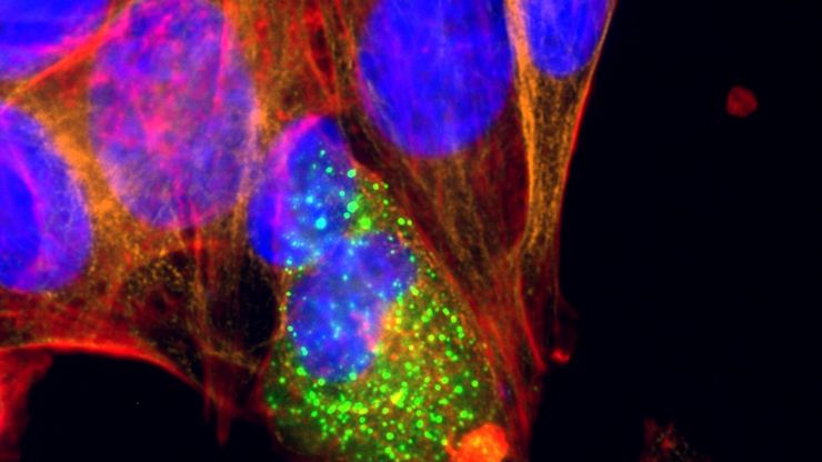
This eBook explores the integration of microscopy and AI technologies in 2D cell culture workflows. It highlights how traditional imaging methods—such as brightfield, phase contrast, and…
人工智能驱动的乳腺癌研究多重染色成像空间分析工具
乳腺癌(BC)是女性因癌症死亡的主要原因,研究查肿瘤微环境(TME)对于阐明肿瘤进展机制至关重要。利用超多标染色空间蛋白质组学技术系统地绘制肿瘤微环境图谱可以提高精准免疫肿瘤学的能力。在这里,我们将基于人工智能的高倍空间分析应用于BC组织,研究免疫细胞类型和生物标记物,从而深入了解受免疫疗法反应的TME分子机制。
利用光片显微技术聚焦三维长时程成像
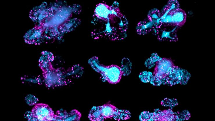
长时程三维成像揭示了复杂的多细胞系统是如何生长和发育的,以及细胞是如何随着时间的推移而移动和相互作用的,从而揭示了发育、疾病和再生方面的重要知识。光片显微镜一次只照射样品的一个薄片,大大减少了光损伤,保护了样品的活性。这种温和的高速技术可在数小时甚至数天内提供清晰的体数据,使研究人员能够实时捕捉生物学的发展过程。
Ultramicrotome Sectioning of Polymers for TEM Analysis
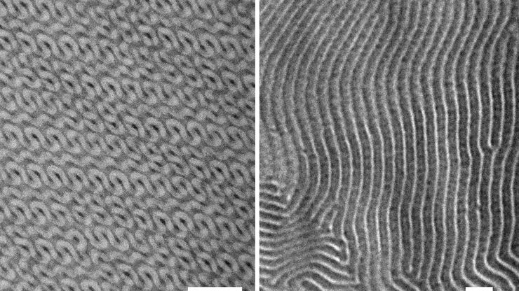
We demonstrate the capabilities of the UC Enuity ultramicrotome from Leica Microsystems for preparing ultrathin sections of polymer samples under both ambient and cryogenic conditions. By presenting…
Biomarker Discovery with Laser Microdissection
Explore the potential of spatial proteomics workflows, such as Deep Visual Proteomics (DVP), to decipher pathology mechanisms and uncover druggable targets.
Altered protein expression, abundance, or…
Volume EM and AI Image Analysis
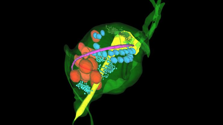
The article outlines a detailed workflow for studying biological tissues in three dimensions using volume-scanning electron microscopy (volume-SEM) combined with AI-assisted image analysis. The focus…
空间蛋白质组学的突破如何拯救生命
中毒性表皮坏死溶解症(TEN)是一种罕见的、但对抗生素或痛风治疗等常见药物的破坏性反应。这种疾病开始时并无大碍,通常只是皮疹,但会迅速升级为大面积皮肤脱落,类似于严重烧伤。尽管 TEN病情十分严重,但其基本机制仍然难以捉摸,治疗方案也仅限于支持性护理。TEN 的死亡率高达 30%,长期以来一直是临床医生的噩梦,直到现在才有了靶向疗法。
A Guide to C. elegans Research – Working with Nematodes
Efficient microscopy techniques for C. elegans research are outlined in this guide. As a widely used model organism with about 70% gene homology to humans, the nematode Caenorhabditis elegans (also…

