
-b-poly(isoprene). Right: Poly(styrene)-b-poly(methyl methacrylate).")
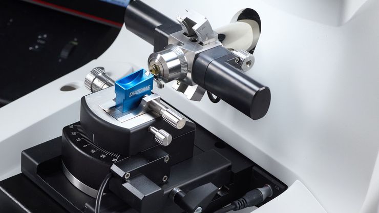
聚合物透射电镜分析用超薄切片技术
本文全面展示了徕卡UC Enuity超薄切片机在聚合物样品超薄切片制备中的优异表现,无论是常温还是低温环境,它都能提供理想的分析样本。文中展示的高分辨率二维及三维TEM图像,有力印证了该仪器在聚合物结构分析领域,对于获得精确、可重复的样品制备结果不可或缺。

体电子显微学与人工智能图像分析
该文章详细阐述了利用体电子显微镜技术 (volume-SEM) 结合人工智能辅助图像分析,对生物组织进行三维研究的工作流程。研究的重点是一种名为毛滴虫的原生动物,这是一种有鞭毛的寄生虫,是导致性传播感染——滴虫病的病原体。为了可视化其复杂的内部结构,研究人员采用了体电子显微镜技术,通过对一系列超薄切片进行成像来重建三维模型。

用于三维生物成像的集成连续切片与冷冻电镜工作流程
本场网络研讨会探讨了集成化工具如何支持从样品制备到图像分析的电子显微镜全流程。专家Andreia Pinto博士、Adrian Boey博士与Hoyin Lai博士将介绍UC Enuity超薄切片机和Aivia图像分析平台,并演示这些工具如何同时适用于常温与低温实验环境。会议内容包含阵列断层成像、基于深度学习的图像分割、以及生物成像中cryo-lift-out工作流程的实际案例解析。

通过Cryo-EM(冷冻电镜)和 CryoFIB(冷冻聚焦离子束) 揭示钠电池退化机制
探索低温电镜和聚焦离子束技术如何揭示钠电池界面的内在结构。本次研讨会将提出基于隔膜渗透(而非枝晶生长)的新型退化模型,并解析电解液溶剂如何影响界面稳定性与电池性能。

"Waffle方法":使用高压冷冻制备复杂样品
本文介绍了一种特殊的高压冷冻方法,即 "Waffle 方法 "的优点。了解 "Waffle 方法 "如何使用电镜载网作为高压冷冻的载体,从而减少样本厚度并支持复杂生物样本的高效低温电子显微镜工作流程。此外,本文还强调了现代 HPF 系统-徕卡微系统公司 EM ICE 的优势,并列举了 EM ICE 用于 "Waffle方法 "的参考文献。
与Helmut Gnaegi一起掌握聚合物超薄切片技术
说到超薄切片技术,很少有人能像Helmut Gnaegi这样举足轻重。作为全球领先的金刚石切片刀公司Diatome的联合创始人,Helmut花了数十年时间完善切片的艺术和科学。在这次独家专访中,他分享了自己在聚合物切片方面的深厚专业知识--从刀具几何形状的细微差别到低温技术的挑战。无论您是经验丰富的电镜专家,还是刚刚起步,Helmut的见解都能为您提供实用的指导和灵感,帮助您获得完美切片。
。")
超薄切片树脂内荧光技术方案
电子显微镜,包括透射电子显微镜 (TEM) 和扫描电子显微镜 (SEM),被广泛应用于获取生物样本或非生物材料的精细结构信息。超薄切片技术是制备厚度小于100纳米的超薄切片的首选方法,适用于透射电镜/扫描电镜分析。样品制备过程中,微小样本块被包埋于环氧或丙烯酸树脂中,去除多余树脂后,使用玻璃刀或金刚石刀将标本切成超薄切片 (50 nm - 100 nm)。

如何通过自动化超薄切片技术节省时间与样本
本文阐述了如何利用树脂包埋电镜样本的 3D micro-CT 数据,在切片前将样本修整至预设目标平面。采用Leica UC Enuity 系统的交互式自动化方案,可显著节省时间、减少样本损耗及缩短新手用户的培训周期。
 and oblique (right) brightfield illumination using a Leica compound microscope. The defect on the wafer surface is clearly more visible with oblique illuminati")
显微镜不同观察方法下的快速半导体检测
已刻蚀的晶圆和集成电路(IC)在生产过程中的半导体检测对于识别和减少缺陷非常重要。
