EM GP2
电子显微镜样品制备
产品
首页
Leica Microsystems
EM GP2 自动投入冷冻仪
重现性和样本质量
阅读我们的最新文章
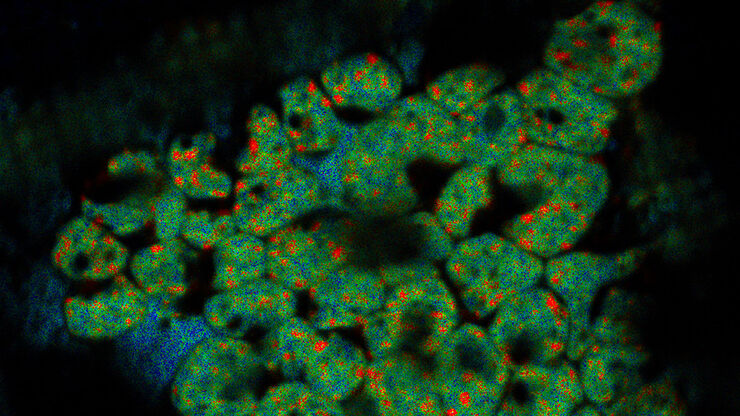
 labeled with membrane-permeable calcein, high-pressure frozen in salt water using EM ICE.")
High-Pressure Freezing for Organoids: Cryo CLEM & FIB Lift Out
Master cryo EM workflow steps for challenging 3D samples: when to choose HPF vs. plunge freezing, reproducible blotting/ice control, contamination aware transfers, Cryo CLEM 3D targeting in organoids,…

从显微镜到电镜:完整的冷冻光电联用工作流程
在题为“多模态玻璃化征程,从实验台到电子显微镜的冷冻关联工作流程”的网络研讨会上,专家团队(Edoardo D'Imprima、Zhengyi Yang、Andreia Pinto 和 Martin…
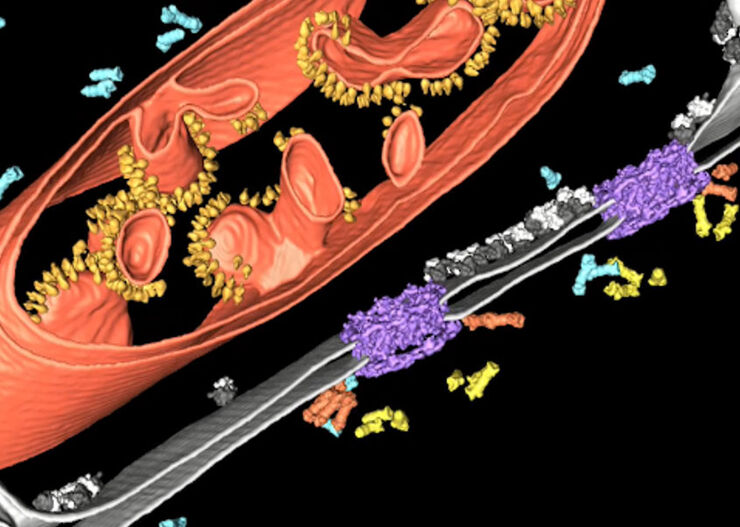
冷冻电子断层扫描
冷冻电子断层扫描(CryoET)用于分辨细胞环境内的生物分子,分辨率达到前所未有的一纳米以下。

冷冻电子显微镜的工作流程与仪器配置
冷冻电子显微镜作为一种日益流行的研究大分子复合体结构的模态,已在细胞生物学领域促成了众多新见解。近年来,该技术进一步向原位结构生物学方向拓展,成为解析自然状态下结构的首选技术。同样,冷冻断裂与冷冻扫描电子显微镜(SEM)的应用也日益广泛。
病毒学
您的主要研究对象是病毒感染和疾病吗? 了解如何使用徕卡显微系统公司的成像和样本制备解决方案深入研究病毒学。
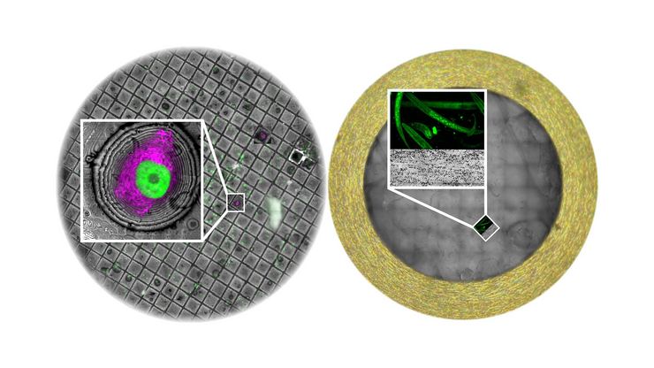
改善冷冻电子断层扫描工作流程
徕卡显微系统有限公司和赛默飞世尔科技有限公司合作开发了一个整条技术路线的冷冻电子断层扫描工作流程。它确保从通过THUNDER成像仪EM冷冻CLEM(也可选择新版的CORAL Cryo冷冻共聚焦CLEM)预选与我们的EM GP2的玻璃化冷冻到Thermo Scientific Krios™ G3i Cryo TEM的3D图像重建的完全整合。所有仪器之间的无缝通信能够获得可靠的结果和可重现的实验。

专家在低温扫描电镜工作流程高压冷冻和冷冻断裂方面的知识
深入了解实验室工作方法并了解在EM样本制备过程中低温扫描电镜研究的优势。了解如何将高压冷冻、冷冻断裂和冷冻传送添加到低温扫描电镜工作流程中,以及徕卡组合如何确保这些不同步骤之间的兼容性。

冷冻透射电子显微镜的投入式冷冻技术:应用
低温下观察完全含水、未染色样本的透射电子显微镜(cryo TEM)是结构生物学、细胞生物学、药理学和其他科学分支的通用工具。通过将标本放入冷冻剂中进行超快速冷冻(投入式冻结)是一种常用的方法,用于制备在透射电镜观察的各种标本。本文是对投入式冷冻的补充,介绍了在不同领域使用投入式冷冻标本的三种冷冻TEM应用。
电子显微镜样品制备工作流程& 用途
通过使用徕卡样品制备解决方案,研究人员可以在利用电子显微镜对样品进行成像时,始终获得高质量、精确且可重复的结果。我们的解决方案支持扫描电子显微镜(SEM)、透射电子显微镜(TEM)及冷冻电镜(Cryo EM)。
应用领域
电子显微镜样品制备工作流程& 用途
通过使用徕卡样品制备解决方案,研究人员可以在利用电子显微镜对样品进行成像时,始终获得高质量、精确且可重复的结果。我们的解决方案支持扫描电子显微镜(SEM)、透射电子显微镜(TEM)及冷冻电镜(Cryo EM)。
