Webinars

最新市场活动资讯

全部活动

12 Nov 2025
12 Nov 2025
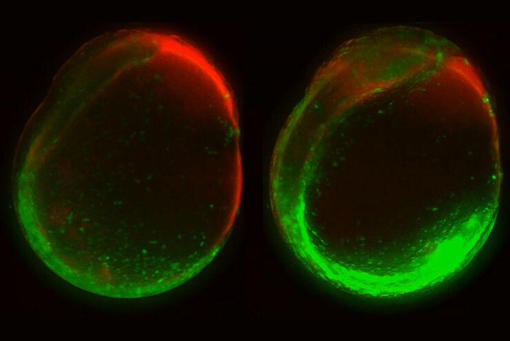
Developing zebrafish (Danio rerio) embryo, from sphere stage to somite stages.

研究斑马鱼胚胎的早期发育阶段

第2集MicaCam的内容是结合宽场和共聚焦成像来研究斑马鱼胚胎(Danio rerio)的早期发育阶段,即从卵细胞到多细胞阶段。
U2OS cells stained with Hoechst for nuclei (blue), MitoTracker green (Mitochondria structure, green) and TMRE (active mitochondria, magenta) and SiR for tubulin (red). Simultaneous acquisition of four channel large area overview using Spiral scan feature using the 10x/1.20 CS2 Water MotCORR objective.

如何获得具有完全时空相关性的多标记实验数据

首期MicaCam会聚焦于活细胞实验当中的挑战。我们的主持人Lynne Turnbull和Oliver Schlicker将以活细胞内线粒体活动研究为例,手把手为您展示如何用多孔板培养箱设计您的实验,以及如何分析结果。
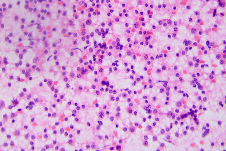
Acute lymphoblastic leukemia

如何从数字细胞病理学中获益

如果您认为数字细胞病理学的特征在于玻璃载玻片的数字化,那么意大利萨勒诺大学医院亚历山德罗·卡普托博士(Alessandro Caputo)的这场网络研讨会将为您拓宽视野。您可以深入了解如果在整个实验室工作流程中采用数字技术,将会实现哪些可能性。
Separation of cells based on their tracking status: A colourised binary mask of a time-lapse microscopy field of view of medium confluency with individual cells highlighted as survivors if they can be tracked since the initial movie frame (cyan), incomers if they migrated into the field of view throughout the movie (yellow) or mistracks if an error occurred in the automated trajectory reconstruction (red).

使用深度学习技术追踪单细胞

人工智能解决方案在显微镜领域的应用不断拓展。从自动化目标分类到虚拟染色,机器学习和深度学习技术在帮助显微镜学家简化分析工作的同时,也在持续推动科学技术领域的突破。
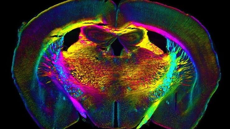
Analysis of anatomy and axon orientation of an adult mouse brain tissue with QLIPP.

通过光学属性了解细胞结构

在过去3年中,显微学家开始在广泛的应用中使用人工智能解决方案,包括图像采集优化(智能显微镜)、目标分类、图像分类、分割、还原、超分辨率和虚拟染色。

如何使用数码显微镜简化检验流程

在“如何使用数码显微镜简化检验流程”的网络研讨会上,他深入探讨了如何使用Emspira 3数码显微镜改进目视检查流程。
Scroll to top