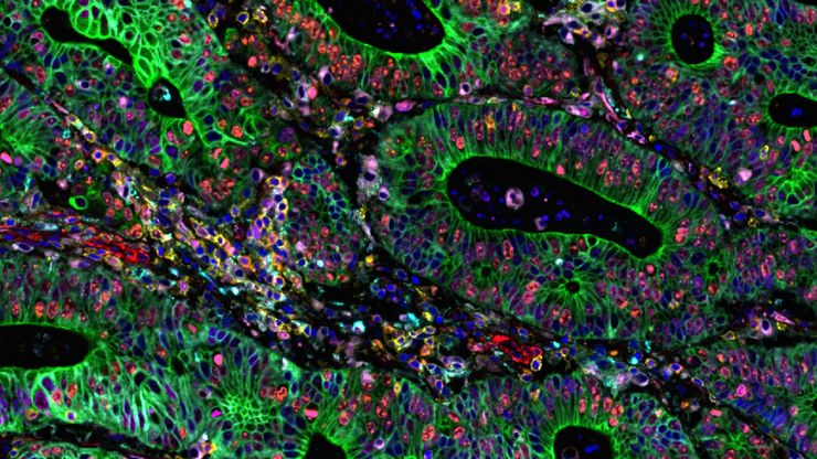
人工智能驱动的乳腺癌研究多重染色成像空间分析工具
乳腺癌(BC)是女性因癌症死亡的主要原因,研究查肿瘤微环境(TME)对于阐明肿瘤进展机制至关重要。利用超多标染色空间蛋白质组学技术系统地绘制肿瘤微环境图谱可以提高精准免疫肿瘤学的能力。在这里,我们将基于人工智能的高倍空间分析应用于BC组织,研究免疫细胞类型和生物标记物,从而深入了解受免疫疗法反应的TME分子机制。
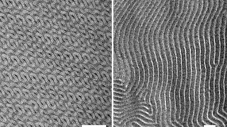
Ultramicrotome Sectioning of Polymers for TEM Analysis
We demonstrate the capabilities of the UC Enuity ultramicrotome from Leica Microsystems for preparing ultrathin sections of polymer samples under both ambient and cryogenic conditions. By presenting…
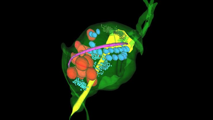
Volume EM and AI Image Analysis
The article outlines a detailed workflow for studying biological tissues in three dimensions using volume-scanning electron microscopy (volume-SEM) combined with AI-assisted image analysis. The focus…
A Novel Laser-Based Method for Studying Optic Nerve Regeneration
Optic nerve regeneration is a major challenge in neurobiology due to the limited self-repair capacity of the mammalian central nervous system (CNS) and the inconsistency of traditional injury models.…
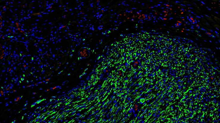
如何为深层肌肉组织中的轴突再生成像
这项研究重点介绍了亚伦-李(Aaron Lee)博士对截肢后肌肉移植中神经再生的定位研究。肢体缺失通常会导致生活质量下降,这不仅是因为组织缺失,还因为轴突再生紊乱引起的神经性疼痛。Mica组织学成像和荧光成像可帮助了解神经再生过程中轴突的生长和分支这项研究有助于塑造未来的神经假体接口设计,改善患者的治疗效果。
Capturing Developmental Dynamics in 3D
This application note showcases how the Viventis Deep dual-view light sheet microscope was successfully used by researchers for exploring high-resolution, long-term imaging of 3D multicellular models…
多重成像揭示结肠癌的肿瘤免疫格局
由于抗药性和复发,癌症免疫疗法获益者寥寥无几,而针对癌症免疫周期多个步骤的组合治疗策略可能会改善治疗效果。这项研究表明,高通量空间蛋白质组学可用于识别细胞生物标志物之间的相互作用,并通过绘制肿瘤免疫微环境图来指导精准的组合疗法。
Improving Zebrafish-Embryo Screening with Fast, High-Contrast Imaging
Discover from this article how screening of transgenic zebrafish embryos is boosted with high-speed, high-contrast imaging using the DM6 B microscope, ensuring accurate targeting for developmental…
超薄切片树脂内荧光技术方案
电子显微镜,包括透射电子显微镜 (TEM) 和扫描电子显微镜 (SEM),被广泛应用于获取生物样本或非生物材料的精细结构信息。超薄切片技术是制备厚度小于100纳米的超薄切片的首选方法,适用于透射电镜/扫描电镜分析。样品制备过程中,微小样本块被包埋于环氧或丙烯酸树脂中,去除多余树脂后,使用玻璃刀或金刚石刀将标本切成超薄切片 (50 nm - 100 nm)。


