联系我们

Leica Microsystems

Leica Microsystems

Leica Microsystems is a world leader in microscopes and scientific instruments. Founded as a family business in the nineteenth century, the company’s history was marked by unparalleled innovation on its way to becoming a global enterprise.

Its historically close cooperation with the scientific community is the key to Leica Microsystems’ tradition of innovation, which draws on users’ ideas and creates solutions tailored to their requirements. At the global level, Leica Microsystems is organized in three divisions, all of which are among the leaders in their respective fields: Life Science, Industry and Medical.

The company is represented in over 100 countries with 6 manufacturing facilities in 5 countries, sales and service organizations in 20 countries, and an international network of dealers. The company is headquartered in Wetzlar, Germany.

http://www.leica-microsystems.com/

Automated Laser Microdissection for Proteome Analysis

组织中的精密空间蛋白质组学信息

尽管可使用基于成像和质谱的方法进行空间蛋白质组学研究,但是图像与单细胞分辨率蛋白丰度测量值的关联仍然是个巨大的挑战。最近引入的一种方法,深层视觉蛋白质组学(DVP),将细胞表型的人工智能图像分析与自动化的单细胞或单核激光显微切割及超高灵敏度的质谱分析结合在了一起。DVP在保留空间背景的同时,将蛋白丰度与复杂的细胞或亚细胞表型关联在一起。
[Translate to chinese:] These images show the microstructure of a hard metal with 10% cobalt which is used for heavy-duty tools. The large increase in magnification of the right image (compared to the left) has a risk of being outside the useful range or, in other words, empty magnification.

当心“无效”放大率

在一个最简化的情况,光学显微镜由一个靠近标本的透镜(物镜)和一个靠近眼睛的透镜(目镜)组成。显微镜放大率是两个显微镜透镜系数的乘积。比如,40倍物镜和10倍目镜可以得到400倍放大率。
Developing embryos of different species at different stages during the elongation of their posterior body axis, from left to right in developmental time. The labelled regions in red depict a region of undifferentiated cells called the tailbud, with the corresponding region generated from that tissue shaded in grey. Upper row: lamprey; middle row: catshark; bottom row, zebrafish. This figure has been adapted from the following publication: Steventon, B., Duarte, F., Lagadec, R., Mazan, S., Nicolas, J.-F., & Hirsinger, E. (2016). Species tailoured contribution of volumetric growth and tissue convergence to posterior body elongation in vertebrates. Development, 2016. 143(10):1732-41

How to Study Gene Regulatory Networks in Embryonic Development

Join Dr. Andrea Boni by attending this on-demand webinar to explore how light-sheet microscopy revolutionizes developmental biology. This advanced imaging technique allows for high-speed, volumetric…
[Translate to chinese:] Multiplexed Cell DIVE imaging of Adult Human Alzheimer’s brain tissue section demonstrating expression of markers specific to astrocytes (GFAP, S100B), microglia (TMEM119, IBA1), AD-associated markers (p-Tau217, β-amyloid) and immune cells such as CD11b+, CD163+, CD4+, and HLA-DRA+, clustered around the β-amyloid plaques.

Spatial Analysis of Neuroimmune Interactions in Alzheimer’s Disease

Alzheimer’s disease (AD) is a complex neurodegenerative disorder characterized by neurofibrillary tangles, β-amyloid plaques, and neuroinflammation. These dysfunctions trigger or are exacerbated by…
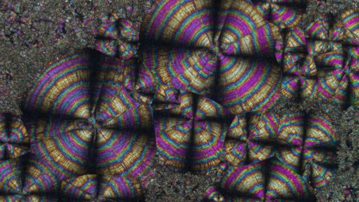
Image of a tartaric-acid crystal taken with polarization microscopy. Tartaric acid, a diprotic, aldaric carboxylic acid, is a naturally occurring organic compound notably found in grapes.

偏振光显微观察

偏光显微镜通常应用于材料科学和地质学领域,根据矿物的折射特性和颜色来识别矿物。在生物学中,偏光显微镜通常用于晶体等双折射结构的识别或成像,或用于植物细胞壁中纤维素和淀粉粒的成像。

A Guide to Spatial Biology

What is spatial biology, and how can researchers leverage its tools to meet the growing demands of biological questions in the post-omics era? This article provides a brief overview of spatial biology…
GLP-1 and PYY localized to distinct secretory pools in L-cells.

Cutting-Edge Imaging Techniques for GPCR Signaling

With this webinar on-demand enhance your pharmacological research with our webinar on GPCR signaling and explore cutting-edge imaging techniques that aim to understand how GPCR signaling translates…
[Translate to chinese:] Salmonella biofilms 3D render

探索微生物世界:三维食品基质中的空间相互作用

Micalis 研究所是与 INRAE、AgroParisTech 和巴黎萨克雷大学合作的联合研究单位。其使命是开发食品微生物学领域的创新研究,以促进健康。在这一系列视频中,Micalis…
[Translate to chinese:] Mouse brain (left) microdissected with a 10x objective (upper right). Inspection of the collection device (lower right).

激光微切割(LMD)促进的分子生物学分析

使用激光微切割(LMD)提取生物分子、蛋白质、核酸、脂质和染色体,以及提取和操作细胞和组织,可以深入了解基因和蛋白质的功能。它在神经生物学、免疫学、发育生物学、细胞生物学和法医学等多个领域有广泛应用,例如癌症和疾病研究、基因改造、分子病理学和生物学。LMD 也有助于研究蛋白质功能、分子机制及其在转导途径中的相互作用。
[Translate to chinese:] Multiplexed Cell DIVE imaging to characterize the spatial landscape in Human Alzheimer’s Cortical Tissue

使用空间多重化探测人类阿尔茨海默病皮层切片

阿尔茨海默病(AD)是最常见的神经退行性疾病,其特征是认知功能的逐渐下降。对 AD 大脑的空间分析可能揭示细胞关系,从而促进对疾病病因的更好理解。本研究捕捉了 AD 皮层组织成分的全球概述,并强调了 Cell DIVE 成像的简化工作流程,从数据采集到使用 Aivia 软件的基于人工智能的分析,最终实现更快的洞察。
[Translate to chinese:] Brightfield image of a pig liver stained with hematoxylin-eosin (HE).

空间代谢组学:探索肿瘤复杂性和治疗见解

在癌症研究中,理解肿瘤细胞与其微环境之间的相互作用至关重要,因为肿瘤微环境显著影响肿瘤进展。空间代谢组学是一种由研究人员开发的新方法,用于研究这一复杂性。通过揭示肿瘤微环境中的空间变化,该方法提供了对其多样化成分及其组织的宝贵见解。这些见解不仅影响临床结果,还为治疗反应提供信息,为个性化治疗策略铺平道路。
[Translate to chinese:] Immunofluorescence image of a mouse enodmetrial organoid stained with CK14 and DAPI

通过子宫内膜类器官推进子宫再生疗法

姜教授的团队研究决定胚胎植入和妊娠成功维持的子宫微环境的重要因素。他们正在开发新的治疗策略,以再生子宫内膜功能,帮助患有子宫内膜疾病(如阿什曼综合症)的患者。她的团队将 3D 子宫内膜类器官移植到小鼠模型中,以识别子宫的显著再生能力背后的细胞和分子机制。从这次访谈中了解她的团队正在进行什么样的研究,以及Mica是如何提供帮助的。
[Translate to chinese:] Mosaic scan of a Masson-Goldner stained cat brain. Magnification: 20x.

基于激光显微切割的稀疏细胞脂质组学分析

通过高覆盖率靶向脂质组学分析稀疏细胞,深入探讨细胞复杂性。这种先进的方法结合了激光显微切割(LMD)和液相色谱-质谱/质谱(LC-MS/MS),揭示了单细胞水平的代谢变化,阐明了糖尿病和肥胖等疾病。通过采用激光显微切割(LMD)获得无污染样本,并使用 SCIEX 7500 系统提高灵敏度,该方法成功检测到 285…
[Translate to chinese:] Image of magnetic steel taken with a 100x objective using Kerr microscopy. The magnetic domains in the grains appear in the image with lighter and darker patterns. A few domains are marked with red arrows. Courtesy of Florian Lang-Melzian, Robert Bosch GmbH, Germany.

使用克尔显微镜快速可视化钢中的磁畴

磁性材料中磁畴与偏振光相互作用后光的旋转,称为克尔效应,使得使用克尔显微镜对磁化样品进行研究成为可能。它可以快速可视化材料表面的磁域。对于用于电气和电子设备的磁性材料(例如钢合金)的高效研发和质量控制,克尔显微镜可以发挥重要作用。本文详细描述了如何使用克尔显微镜对钢合金晶粒中的磁域进行成像。
[Translate to chinese:] AI-based cell counting performed with a phase-contrast and fluorescence image using the Mateo FL microscope.

利用AI增强的细胞计数实现精准和高效

本文描述了利用AI进行精确和高效的细胞计数。准确的细胞计数对于 2D 细胞培养的研究至关重要,例如细胞动力学、药物发现和疾病建模。精确的细胞计数对于确定细胞存活率、增殖速率和实验条件的影响至关重要。这些因素对于可靠和稳健的结果至关重要。描述了基于人工智能的方法如何显著提高细胞计数的准确性和速度,从而对细胞研究产生重大影响。
[Translate to chinese:] AI-based transfection analysis (left) of U2OS cells which were transfected with a fluorescently labelled protein. A fluorescence image of the cells (right) is also shown. The analysis and imaging were performed with Mateo FL.

利用AI实现细胞转染的高效分析

本文探讨了AI(AI)在优化 2D 细胞培养研究中转染效率测量中的关键作用。对于理解细胞机制而言,精确可靠的 2D 细胞培养转染效率测量至关重要。靶向蛋白的高转染效率对于包括活细胞成像和蛋白纯化在内的实验至关重要。手动估计存在不一致性和不可靠性。借助AI的力量,可以实现高效可靠的转染研究。
[Translate to chinese:] Image of confluent cells taken with phase contrast (left) and analyzed for confluency using AI (right).

通过 AI 汇合度提高 2D 细胞培养的精度

本文解释了如何利用人工智能(AI)进行高效、精确的 2D 细胞培养汇合度评估。准确评估细胞培养的汇合度,即表面积覆盖的百分比,对于可靠的细胞研究至关重要。传统方法使用视觉检查或简单算法,使结果不客观和精确,尤其是对于用于药物发现、组织工程和再生医学的复杂细胞系。利用自动化图像分析和深度学习算法的方法提供更好的精度,并可以增强实验结果。
Cell DIVE image of stromal remodeling around B cell follicles of follicular lymphoma patients. Stromal cells labeled with antibodies against desmin (red), SPARC (orange), vimentin (blue), and a-sma (yellow). Extracellular matrix labeled with antibody against lumican (cyan). B cells labeled with antibody against CD20 (green). Image credit: Dr. Andrea Radtke, Center for Advanced Tissue Imaging, NIAID, NIH

Cell DIVE开放式超多重免疫荧光成像如何赋能空间生物学

空间生物学和多重成像工作流程在免疫肿瘤学研究中变得越来越重要。许多研究人员即使使用有效的工具和方案,也很难提高研究效率。我们将介绍研究人员如何利用开放式超多重免疫荧光的适应性,将 IBEX 成像与Cell DIVE 相结合,创造了一种名为 Cell DIVE-IBEX 的技术。它让这些研究人员能够调整现有的技术和试剂,并获得Cell DIVE 在其免疫肿瘤学研究中的可扩展性。
[Translate to chinese:] Image of murine dopaminergic neurons which have been marked for laser microdissection (LMD).

利用激光显微切割(LMD)在空间背景下分离神经元

在阿尔茨海默病之后,帕金森病是第二常见的进行性神经退行性疾病。在首发症状出现之前,中脑中高达70%的多巴胺释放神经元已经死亡。本文描述了如何使用现代激光显微切割(LMD)方法帮助解决帕金森病之谜。研究涉及在空间背景下分离和分析神经元。这些细胞来自帕金森病患者的死后黑质组织样本,以便深入了解该病的分子机制。

激光显微切割技术如何助力神经科学研究取得开创性进展?

玛尔塔·帕特林尼博士,卡罗林斯卡学院的高级科学家,分享了她在成人人类神经发生开创性研究中使用激光显微切割(LMD)的经验,并提供了关于LMD在空间蛋白质组学和精准医学中未来应用潜力的个人见解。
Region of a patterned wafer inspected using optical microscopy and automated and reproducible DIC (differential interference contrast). With DIC users are able to visualize small height differences on the wafer surface more easily.

6英寸晶圆检测显微镜:可靠观察细微高度差异

本文介绍了一种配备自动化和可重复的DIC(微分干涉对比)成像的6英寸晶圆检测显微镜,无论用户的技能水平如何。制造集成电路(IC)芯片和半导体组件需要进行晶圆检测,以验证是否存在影响性能的缺陷。这种检测通常使用光学显微镜进行质量控制、故障分析和研发。为了有效地可视化晶圆上结构之间的小高度差异,可以使用DIC。
[Translate to chinese:] Optical microscope image, which is a composition of both brightfield and fluorescence illumination, showing organic contamination on a wafer surface. The inset images in the upper left corner show the brightfield image (above) and fluorescence image (below with dark background).

晶圆上的光刻胶残留和有机污染物的可视化

随着半导体上集成电路(IC)的尺寸低于10纳米,在晶圆检测中有效检测光刻胶残留等有机污染物和缺陷变得越来越重要。光学显微镜仍然是常见的检测方法,但对于有机污染物,明场和其他类型的照明可能会存在局限性。本文讨论了荧光显微镜如何在半导体行业的QC、故障分析和研发过程中有效检测晶圆上的光刻胶残留和其他有机污染物。
[Translate to chinese:] Evolved ARveo 8: Operating Room (OR) set-up.

增强现实:改变神经外科手术

在这本电子书中,您将探索增强现实(AR)为神经外科领域带来的激动人心的进步。这本综合指南包括解释性视频,解答关键问题并提供详细解释,揭示了外科手术的未来。

利用AI驱动的多重图像分析探索结肠腺癌

在这项研究中,我们展示了一种利用Cell DIVE和AIVIA软件的空间生物学工作流程,以绘制结肠腺癌中的肿瘤免疫景观图。

激光显微切割技术用于组织和细胞分离的协议 - 免费下载电子书

激光显微切割(LMD,也称为激光捕获显微切割或LCM)使用户能够分离特定的单个细胞或整个组织区域,甚至亚细胞结构如染色体。纯化的组织和细胞可用于下游的RNA、DNA和蛋白质组工作流程。
[Translate to chinese:] The role of extracellular signalling mechanisms in the correct development of the human brain

在神经发育过程中,细胞是如何相互交流的?

细胞间通信是大脑发育过程中一个必不可少的过程,它受到多种因素的影响,包括细胞的形态、粘附分子、局部细胞外基质和分泌囊泡。在本次网络研讨会上,您将了解到对这些机制更深入的理解是如何推动对神经发育障碍的理解的。
Intestinal organoids label with FUCCI reporter to follow cell cycle dynamics. Courtesy of Franziska Moos. Liberali lab. FMI Basel (Switzerland).

Dual-View LightSheet Microscope for Large Multicellular Systems

Visualizing the dynamics of complex multicellular systems is a fundamental goal in biology. To address the challenges of live imaging over large spatiotemporal scales, Franziska Moos et. al. present…

肿瘤空间微环境的元癌症分析

研究 TME中肿瘤、基质和免疫细胞之间的相互作用需要采用超多重免疫荧光成像方法。在这里,我们分析了一组Cell Signaling Technology(CST®)抗体,这些抗体针对肺癌、结肠癌和胰腺癌等癌症的标志物。通过Cell DIVE成像和Aivia中的聚类分析,我们确定了TME中的空间相互作用,包括组织特异性和共有的相互作用。
[Translate to chinese:] Multiplexed Cell DIVE imaging of Colon Adenocarcinoma (CAC) tissue. A panel of approximately 30 biomarkers targeted towards various leukocyte lineages, epithelial, stromal, and endothelial cell types was utilized to characterize the tumor immune microenvironment in human colon adenocarcinoma (CAC) tissue.

通过成像和AI绘制结直肠癌的景观

结肠癌是一种高负担疾病。尽管进行了化疗干预和手术切除,但疾病可能会复发。了解结肠癌微环境对于改善治疗效果是必要的。在这里,我们使用空间生物学方法,通过Cell DIVE和 Aivia可视化结肠腺癌组织中的30个生物标志物。我们探讨了肿瘤组织的血管化、免疫细胞反应和细胞增殖。
Clustering based analysis reveals various immune cell populations enriched in tumor cells within CT26.WT syngeneic mouse tumor models.

肿瘤组织中肿瘤和免疫细胞的空间结构

免疫检查点阻断(ICB)疗法在许多癌症中具有临床益处,但一些患者并无反应。最佳的治疗组合可能受到肿瘤内存在的免疫抑制机制的影响。
Scroll to top