工业

工业

工业

深入探讨有关工业和病理学领域的高效检测、优化工作流程和提高人体工学舒适度的文章和网络研讨会。涉及的主题包括质量控制、材料分析、病理学显微镜等。在这里您可以获得有关使用前沿技术提高生产力和优化质量以及准确地进行病理学诊断的干货。
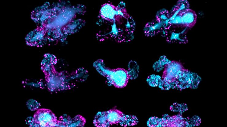
67-hour, multi-position time-lapse of mouse intestinal organoids expressing the cell cycle reporter FUCCI2 (hGem-mVenus and hCdt1-mCherry).

利用光片显微技术聚焦三维长时程成像

长时程三维成像揭示了复杂的多细胞系统是如何生长和发育的,以及细胞是如何随着时间的推移而移动和相互作用的,从而揭示了发育、疾病和再生方面的重要知识。光片显微镜一次只照射样品的一个薄片,大大减少了光损伤,保护了样品的活性。这种温和的高速技术可在数小时甚至数天内提供清晰的体数据,使研究人员能够实时捕捉生物学的发展过程。
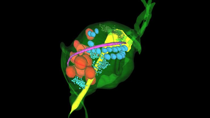
Final Segmentation of organelles in Trichomonas species. Magenta – costa, light blue – hydrogenosomes, turquoise – ER, red – vacuoles, yellow – axostyle, green – Golgi apparatus.  Sample courtesy of Isabelle Guerin-Bonne, Low Kay En, Electron Microscopy Unit, Yong Loo Lin School of Medicine, National University of Singapore. Scale bar: 1 µm.

Volume EM and AI Image Analysis

The article outlines a detailed workflow for studying biological tissues in three dimensions using volume-scanning electron microscopy (volume-SEM) combined with AI-assisted image analysis. The focus…
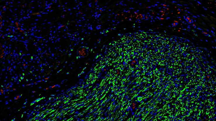
荧光显微镜下的组织切片,显示成年大鼠趾长伸肌与腓总神经之间的界面。2 周后的再生周围神经界面(RPNI)。使用 Mica 采集的图像。细胞核(蓝色)、神经丝(绿色)和 S100B(红色)染色。图片由伦敦帝国学院生物工程系 Aaron Lee 博士(Rylie Green 博士实验室)提供。

如何为深层肌肉组织中的轴突再生成像

这项研究重点介绍了亚伦-李(Aaron Lee)博士对截肢后肌肉移植中神经再生的定位研究。肢体缺失通常会导致生活质量下降,这不仅是因为组织缺失,还因为轴突再生紊乱引起的神经性疼痛。Mica组织学成像和荧光成像可帮助了解神经再生过程中轴突的生长和分支这项研究有助于塑造未来的神经假体接口设计,改善患者的治疗效果。
5 hour time-lapse maximum intensity projection of a zebrafish embryo along the z-axis at 3 days post fertilization. Left: microglia cells. Right: bright field channel. Courtesy of Prof. Francesca Peri, University of Zurich, Switzerland.

Capturing Developmental Dynamics in 3D

This application note showcases how the Viventis Deep dual-view light sheet microscope was successfully used by researchers for exploring high-resolution, long-term imaging of 3D multicellular models…
Pancreatic Ductal Adenocarcinoma with 11 Apoptosis biomarkers shown – BAK, BAX, BCL2, BCLXL, Caspase9, CIAP1, NaKATPase, PCK26, SMAC, Vimentin, and XIAP.

Transforming Research with Spatial Proteomics Workflows

Spatial Proteomics, Nature Methods 2024 Method of the Year, is driving research advancements in cancer, immunology, and beyond. By combining positional data with high throughput imaging of proteins in…
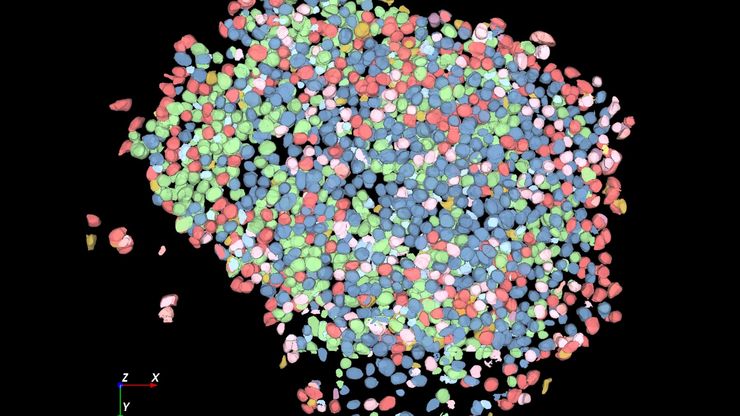
Large volume computational clearing processed Thunder image of human pancreatic islet organoid. Cells segmented using Segment By Example tool, automatically phenotyped, and color-coded based on phenotypes in Aivia. Image courtesy of the Matthias von Herrath Lab, La Jolla Institute of Immunology, La Jolla, CA.

利用人工智能图像分析工具更快、更轻松地获得洞察力

了解 Aivia 如何通过快速设置、准确的人工智能检测和简便的批量处理功能,帮助科学家简化图像分析。
Image: Human stem cell-derived mid brain organoids. Courtesy of Dr Tanya Singh, University of Oxford.

揭开类器官模型在生物医学研究中的秘密

准备深入了解类器官和3D培养物的世界,它们是促进我们了解人类健康的重要工具。浏览这些复杂的结构并获取清晰的图像进行分析是一项挑战。在本次活动中,来自牛津大学和伦敦大学学院的研究人员将与我们一起展示Thunder Imager Cell转盘共聚焦系统 如何提供更有说服力的高质量数据,以便深入了解各种模型。
Transfection using the Uncommon Bio reprogramming system. Image acquired using the THUNDER Imager 3D Cell Culture with THUNDER Large Volume Computational Clearing (LVCC) applied. Image courtesy of Samuel East, Uncommon Bio.

利用新型可扩展的干细胞培养设计未来

具有远见卓识的生物技术初创企业 Uncommon Bio 正在应对世界上最大的健康挑战之一:食品可持续性。在这次网络研讨会上,干细胞科学家塞缪尔-伊斯特(Samuel East)将展示他们如何使细胞农业的干细胞培养基既安全又经济可行。了解他们如何将培养基成本降低 1000 倍,并开发出不含动物成分、食品安全的 iPSC 培养基。
3D culture of ovarian cancer cells imaged using the confocal mode of Mica.

Mica: 助力伦敦帝国学院开展跨学科科研研究

这篇访谈重点介绍了伦敦帝国学院的 Mica 所产生的变革性影响。科学家们解释了Mica如何改变了游戏规则,扩大了研究的可能性,促进了跨学科合作。他们解释了使用 Mica 进行详细的活细胞成像如何提供更有意义的信息,使科学家始终站在研究的最前沿。研究小组预计,Mica将继续开辟新的研究途径,包括研究微流体技术和其他先进应用。
Scroll to top