EnFocus
手术显微镜
产品
首页
Leica Microsystems
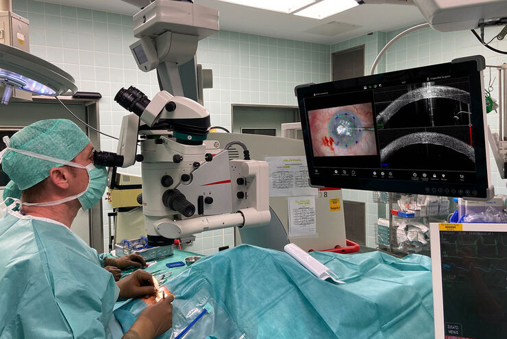
EnFocus 术中 OCT 成像系统
至臻追求
阅读我们的最新文章

眼科案例分析:角膜移植
内皮角膜移植术是一种现代角膜移植技术。角膜移植手术有多种手术方法,包括角膜后弹力膜撕除角膜内皮移植术(DSEK)和角膜后弹力膜内皮移植术(DMEK)。这些方法在植入供体组织的数量上有所不同。

术中OCT引导的青光眼支架修复手术
青光眼是导致全球不可逆失明的主要原因之一。小梁网切除术和导管分流引流术等历史悠久的手术技术会带来巨大的短期风险和潜在并发症。近年来,随着微创青光眼手术(MIGS)的出现,手术方法有了长足的发展,其特点是对组织的破坏最小、内路粘小管植入、手术时间短、器械简单、术后恢复快。

实时 OCT 成像如何影响角膜手术的精确性?
角膜手术是一个高度专业化的领域。它需要极高的显微精确度,以克服诸如清晰可视化整个前房、进行后弹力层剥离和内皮手术、测量切割角膜基质的深度以及量化切口深度,同时避免对供体组织造成损伤等挑战。请观看丰塔纳教授如何在角膜移植手术中使用实时术中光学相干断层扫描成像来提高他的手术精确度。

RPE65 基因治疗视网膜下注射:术中 OCT 的优点
术中 OCT 如何支持 RPE65 基因治疗视网膜下注射程序以监测视网膜下滤泡、中心凹拉伸和视网膜切开术闭合。

术中 OCT 辅助下的脱位性白内障继发房角闭合病例
了解如何在术中 OCT 的辅助下通过闭角术治疗脱位白内障,从而获得长期的良好效果,避免日后晶状体脱位。

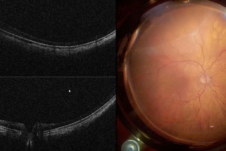
后节手术: 利用术中OCT技术的好处
术中光学相干断层扫描 (OCT) 支持后段病变的精确定位、评估和管理,在后段和玻璃体手术中提供先进的可视化和洞察力。

术中OCT辅助角膜移植手术
了解术中OCT如何在角膜移植手术中为角膜外科医生提供更佳的可视化支持,并促进供体角膜的定位和适应

术中光学显像如何帮助青光眼手术获得更多洞察力
青光眼是导致全球失明的主要原因之一。青光眼手术可以延缓疾病的发展。在青光眼手术过程中,术中光学相干断层扫描(OCT)的使用为眼科外科医生提供了更佳的可视化效果,让他们更深入地了解表面下组织对手术操作的反应。 莫拉鲁博士通过两个原发性开角型青光眼(POAG)的临床病例强调了它的价值。

眼科: 复杂白内障手术中的可视化
白内障手术是最常见的眼科手术。为了满足白内障手术的需要,Ozana Moraru 博士使用了 Leica Microsystems 的 M844 显微镜和 EnFocus 术中光学相干断层扫描 (OCT) 以及 3D 可视化系统。在本案例研究中,她介绍了术中光学相干断层扫描如何为标准和复杂的白内障手术病例提供有用信息。

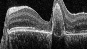
使用光学相干断层扫描改善黄斑孔手术
黄斑裂孔是一种罕见的眼疾,会导致中心视力模糊,影响日常活动。黄斑裂孔通常是由黄斑被牵拉或拉伸而引起的开口,最常见的原因通常是年龄相关的眼部变化。然而,在本病例研究中,Robert A. Sisk博士(医学博士,FACS)介绍了一个小儿眼科病例,其中术中光学相干断层扫描(OCT)为他的手术提供了额外的见解。

眼部基因治疗视网膜下注射
Leber先天性黑矇(LCA)是一组先天性视网膜营养不良,可导致早年出现重度视力损伤。患者通常表现为眼球震颤、瞳孔反应迟钝或近乎缺失、视力严重下降、畏光和高度远视[1]。眼部基因治疗可极大改善这些患者的视力[2]。通过术中视网膜下注射AAV载体完成基因传递。

光学相干断层扫描(OCT)引导下视网膜手术的临床研讨会
在本记录中,来自新加坡某眼科中心的A教授和来自西班牙巴塞罗那某儿童医院的B医生分享了他们使用眼科显微镜所提供的术中OCT行视网膜手术的技术经验。他们报告了从常规黄斑裂孔手术到基因治疗的多个感兴趣儿科病例。
Proveo 8 with intraoperative OCT – a User Evaluation in an University Setting
Optical coherence tomography (OCT) makes structures in the eye visible that lie beneath the surface. When OCT is used intraoperatively, surgeons gain insight into possible pathological changes in the…
术中光学相干断层扫描(OCT)在视网膜手术中的应用
在过去的一年,巴西某视网膜诊所的手术团队采用了显微镜集成式术中OCT。在他们发布的白皮书《术中光学相干断层扫描:视网膜手术的临床经验》中,深入分析了使用OCT的复杂视网膜手术病例,其中OCT额外提供了有用信息,某些情况下这些信息对外科医生做出决策至关重要。

Towards Advanced Use of Intraoperative OCT in Cataract Surgery
In this White Paper, Dr. Rachid Tahiri shares his personal experience with the Leica EnFocus intraoperative OCT, the valuable features supporting smooth surgery and how it allows him to minimize…

眼科手术向常规使用术中OCT的阶段迈进
A博士是最早在眼科手术中使用术中OCT的医生之一。自2016年以来,她累积了大量使用各种不同术中OCT系统的临床经验。在本白皮书中,她对自己的个人经验进行了总结。她解释了为什么她相信术中OCT会继续存在,并在眼科手术实践中有一个坚定的未来。

光学相干断层扫描(OCT)引导下眼科手术如何帮助医生全力聚焦于实现完美
按需观看成功的临床在线研讨会。查看A博士对OCT引导下眼科手术受益的介绍。她报告了临床病例,分享了使用眼科显微镜的组成部分EnFocus术中OCT的经验。
术中OCT可为我们提供什么?
以下视频摘自A博士发布的网络研讨会。在网络研讨会期间,A博士讨论并说明了术中光学相干断层扫描(OCT)如何在复杂的眼前节手术和眼后节手术中提供实时的术中可视信息。她解释了术中OCT如何为显微镜视野提供补充,并为她提供了亚表层组织如何对其手术操作做出反应的即时信息。
眼科OCT成像系统
徕卡眼科OCT成像系统以简单易用的高品质成像技术为眼科医生、眼科主刀医生和研究人员提供支持。
应用领域
眼科
徕卡眼科手术显微镜具有性能卓越的光学器件和照明系统,能帮助术者以高精度完成手术,并改善眼科手术的治疗效果。
视网膜手术
您是否在寻找视网膜手术显微镜解决方案?了解徕卡解决方案的独特优势,以及这些方案可以如何帮助您克服挑战。
角膜手术
Leica Ophthalmic microscopes offer numerous solutions for cornea surgery and support you from corneal donor tissue harvesting and preparation to transplant surgery.
青光眼手术
徕卡眼科显微镜解决方案为小梁切除术、青光眼植入物手术和青光眼微创手术 (MIGS) 提供理想的视觉效果。
