UC Enuity
电子显微镜样品制备
产品
首页
Leica Microsystems
UC Enuity 超薄切片机
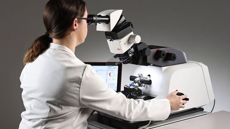
体验先进的自动化和精确度
阅读我们的最新文章
, insulin SGs (orange), microtubules (red), nucleus (yellow), and plasma membrane (transparent).")
High-Pressure Freezing Protocols for Ultrastructural 3D EM
High pressure freezing (HPF) can help preserve hydrated cells and tissues close to their biological state at the moment of immobilization, supporting more reliable ultrastructural interpretation than…
Ultramicrotome UC Enuity in Practice: Stable 15 nm Sections at ZFE
After using the UCT and UC6 ultramicrotomes, Claudia Mayrhofer calls UC Enuity a leap in stability—so robust that vibrations and temperature shifts don’t spoil sections, even with multiple users. Auto…

超薄切片技术电子书:定位、修块& 对刀
超薄切片技术正经历日新月异的发展,当今的显微镜系统对高质量切片、精准定位以及可重复的工作流程提出了更高要求。这本电子书整合了专家应用指南、自动化方法及实操指导,旨在帮助从初学者到资深镜检人员的每一位用户,在电镜、光电联用及体电镜工作流程中,获得一致且可靠的超薄切片。
-b-poly(isoprene). Right: Poly(styrene)-b-poly(methyl methacrylate).")
聚合物透射电镜分析用超薄切片技术
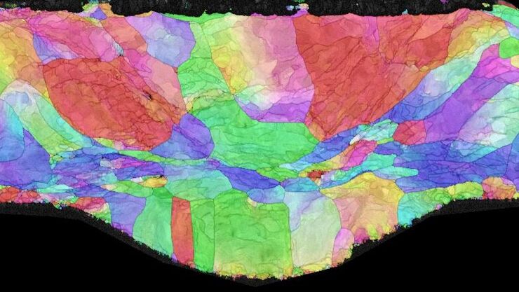
本文全面展示了徕卡UC Enuity超薄切片机在聚合物样品超薄切片制备中的优异表现,无论是常温还是低温环境,它都能提供理想的分析样本。文中展示的高分辨率二维及三维TEM图像,有力印证了该仪器在聚合物结构分析领域,对于获得精确、可重复的样品制备结果不可或缺。

体电子显微学与人工智能图像分析
该文章详细阐述了利用体电子显微镜技术 (volume-SEM) 结合人工智能辅助图像分析,对生物组织进行三维研究的工作流程。研究的重点是一种名为毛滴虫的原生动物,这是一种有鞭毛的寄生虫,是导致性传播感染——滴虫病的病原体。为了可视化其复杂的内部结构,研究人员采用了体电子显微镜技术,通过对一系列超薄切片进行成像来重建三维模型。

用于三维生物成像的集成连续切片与冷冻电镜工作流程
本场网络研讨会探讨了集成化工具如何支持从样品制备到图像分析的电子显微镜全流程。专家Andreia Pinto博士、Adrian Boey博士与Hoyin Lai博士将介绍UC Enuity超薄切片机和Aivia图像分析平台,并演示这些工具如何同时适用于常温与低温实验环境。会议内容包含阵列断层成像、基于深度学习的图像分割、以及生物成像中cryo-lift-out工作流程的实际案例解析。

神经科学研究指南
神经科学通常需要研究具有挑战性的标本,以更好地了解神经系统和疾病。徕卡显微镜帮助神经科学家深入了解神经元功能。
与Helmut Gnaegi一起掌握聚合物超薄切片技术
说到超薄切片技术,很少有人能像Helmut Gnaegi这样举足轻重。作为全球领先的金刚石切片刀公司Diatome的联合创始人,Helmut花了数十年时间完善切片的艺术和科学。在这次独家专访中,他分享了自己在聚合物切片方面的深厚专业知识--从刀具几何形状的细微差别到低温技术的挑战。无论您是经验丰富的电镜专家,还是刚刚起步,Helmut的见解都能为您提供实用的指导和灵感,帮助您获得完美切片。
。")
超薄切片树脂内荧光技术方案
电子显微镜,包括透射电子显微镜 (TEM) 和扫描电子显微镜 (SEM),被广泛应用于获取生物样本或非生物材料的精细结构信息。超薄切片技术是制备厚度小于100纳米的超薄切片的首选方法,适用于透射电镜/扫描电镜分析。样品制备过程中,微小样本块被包埋于环氧或丙烯酸树脂中,去除多余树脂后,使用玻璃刀或金刚石刀将标本切成超薄切片 (50 nm - 100 nm)。

如何通过自动化超薄切片技术节省时间与样本
本文阐述了如何利用树脂包埋电镜样本的 3D micro-CT 数据,在切片前将样本修整至预设目标平面。采用Leica UC Enuity 系统的交互式自动化方案,可显著节省时间、减少样本损耗及缩短新手用户的培训周期。
超薄切片介绍
对样本开展研究时,为了以纳米级分辨率显示其精细结构,通常会使用到电子显微镜。电子显微镜有两种类型:扫描电子显微镜(SEM)用于对样本表面成像,以及需要使用极薄电子透明样本的透射电子显微镜(TEM)。因此,使用电子显微镜对样本内部的精细结构进行成像时,此类技术解决方案需要制作出非常薄的样本切片。被称为超显微技术的样本制备方法可以产生具有最小伪影的超薄切片(厚度20-150nm)。在切片过程中,样本的…

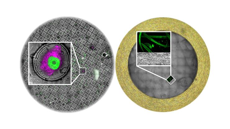
如何在块面中自动获取感兴趣的荧光细胞
本文介绍了使用超薄切片超薄切片机自动修整修块功能,获取树脂块面中带有荧光信号的细胞结构。我们展示了如何使用配置有体视显微镜 M205 FA 的超薄切片超薄切片机 UC Enuity ,来识别感兴趣的荧光细胞,如何自动修整包含细胞的块面,以及如何在切片中观察细胞而无需转移到外部显微镜。

通过自动切片改善您的超薄切片工作流程
在不断发展的电镜样品制备领域,保持领先地位至关重要。这个网络研讨会提供了关于超薄切片最新进展的重要见解,这些进展可以显著增强您实验室的能力。
材料科学样本制备方法的工作流程解决方案
本手册介绍并解释了材料科学样本制备最常用的样本制备方法的工作流程解决方案:

高质量超薄切片:样品与切片刀自动对齐
超薄切片技术是获取样品切片的最常用方法。在室温条件制备时,将样品小块嵌入环氧树脂中,然后通过修剪去除多余的树脂,并使用玻璃刀或金刚石刀将样品切成厚度为50-100纳米之间的薄片。

超薄获得高质量的超薄切片
UC Enuity能够应对这一挑战,提供厚度一致且高质量的切片。其用户友好的设计不仅简化了研究过程,还提高了可重复性,使研究者对所得到的结论更有信心。
 stained with osmium tetroxide (OsO4), sectioned with a DIATOME diamond knife at room temperature, and then imaged with HAADF TEM.")
用于材料切片的超薄切片技术
了解这些用于材料切片的超薄切片技术的宝贵见解,并学习如何克服在处理聚合物、金属和脆性材料时遇到的挑战。千万不要错过这个提高您对先进显微镜样品制备方法的认识的机会。

高级组织成像& 分析
利用徕卡显微系统的先进成像解决方案,深入了解组织结构和功能,从而加深对空间生物学和疾病机制的理解。
电子显微镜样品制备工作流程& 用途
通过使用徕卡样品制备解决方案,研究人员可以在利用电子显微镜对样品进行成像时,始终获得高质量、精确且可重复的结果。我们的解决方案支持扫描电子显微镜(SEM)、透射电子显微镜(TEM)及冷冻电镜(Cryo EM)。
应用领域
生命科学研究
徕卡(Leica)生命科学显微镜凭借先进的创新和专业技术能力,支持观察、测量和分析微结构的成像要求。徕卡显微系统对科学应用领域的高度关注,使徕卡显微系统的用户始终保持领先位置
电子显微镜样品制备工作流程& 用途
通过使用徕卡样品制备解决方案,研究人员可以在利用电子显微镜对样品进行成像时,始终获得高质量、精确且可重复的结果。我们的解决方案支持扫描电子显微镜(SEM)、透射电子显微镜(TEM)及冷冻电镜(Cryo EM)。
高级组织成像& 分析
利用徕卡显微系统的先进成像解决方案,深入了解组织结构和功能,从而加深对空间生物学和疾病机制的理解。
