Cell DIVE
复合光学显微镜
产品
首页
Leica Microsystems
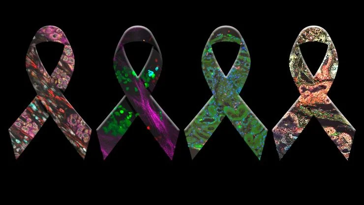
Cell DIVE 超多标组织成像分析整体解决方案
用开放的超多标成像解决方案彻底改变组织研究工作。
阅读我们的最新文章
癌症研究中显微镜的历史、发展和趋势
癌症是一种全球性疾病,2020 年全球将新增 1800 万确诊病例,1000 万人死于癌症。预计到 2040 年,病例数将增加约 55%。研究了解癌症的发生、发展以及开发新的诊断工具和治疗方法至关重要。
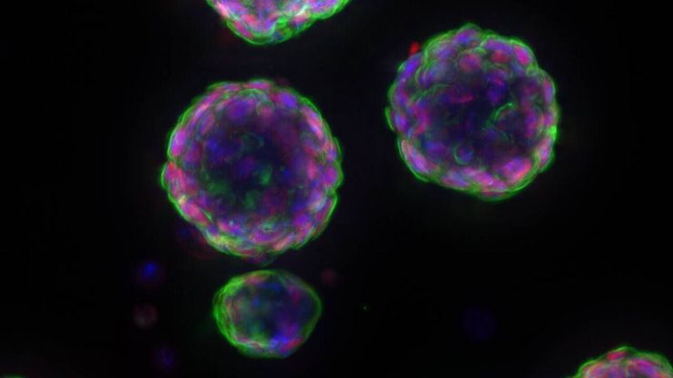
 and astrocytes (green) in a cortical spheroid derived from human induced pluripotent stem cells.")
活细胞成像指南
在生命科学各研究领域的广泛应用中,活细胞成像是一种不可或缺的工具,用于观察细胞在尽可能接近活体(即活的、活跃的)状态下的情况。本指南回顾了确保成功进行活细胞成像的各种重要注意事项,并介绍了各种旨在克服常见挑战的高性能解决方案。这些进展使我们能够对细胞生理学和动力学有新的认识。
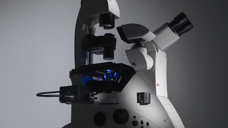
选择研究用显微镜时应考虑的因素
光学显微镜通常是生命科学研究实验室的核心设备之一。它可用于各种应用,揭示许多科学问题。因此,显微镜的配置和功能对其应用范围至关重要,从明视野显微镜到荧光显微镜,再到活细胞成像。本文简要概述了显微镜的相关功能,并总结了在选择研究用显微镜时应考虑的关键问题。
.")
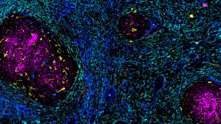
人工智能驱动的乳腺癌研究多重染色成像空间分析工具
乳腺癌(BC)是女性因癌症死亡的主要原因,研究查肿瘤微环境(TME)对于阐明肿瘤进展机制至关重要。利用超多标染色空间蛋白质组学技术系统地绘制肿瘤微环境图谱可以提高精准免疫肿瘤学的能力。在这里,我们将基于人工智能的高倍空间分析应用于BC组织,研究免疫细胞类型和生物标记物,从而深入了解受免疫疗法反应的TME分子机制。

多重成像揭示结肠癌的肿瘤免疫格局
由于抗药性和复发,癌症免疫疗法获益者寥寥无几,而针对癌症免疫周期多个步骤的组合治疗策略可能会改善治疗效果。这项研究表明,高通量空间蛋白质组学可用于识别细胞生物标志物之间的相互作用,并通过绘制肿瘤免疫微环境图来指导精准的组合疗法。

神经科学研究指南
神经科学通常需要研究具有挑战性的标本,以更好地了解神经系统和疾病。徕卡显微镜帮助神经科学家深入了解神经元功能。

波士顿和旧金山创新中心
波士顿和旧金山创新中心将帮助您推动科学发现。我们为研究人员提供最先进的显微镜技术和专家指导。这些中心位于波士顿和旧金山的中心地带,是创新与精准相结合的协作空间。

利用空间蛋白质组学工作流程改革研究工作
空间蛋白质组学是《自然-方法》2024 年度方法,正在推动癌症、免疫学等领域的研究进展。通过将定位数据与组织中蛋白质的高通量成像结合起来,研究人员可以发现疾病进展和治疗反应方面的洞察力,从而更好地了解人类生物学。在这里,您可以了解更多有关空间生物学的信息,以及徕卡显微系统的工具如何推动蛋白质生物标记的可视化和分析取得进展。
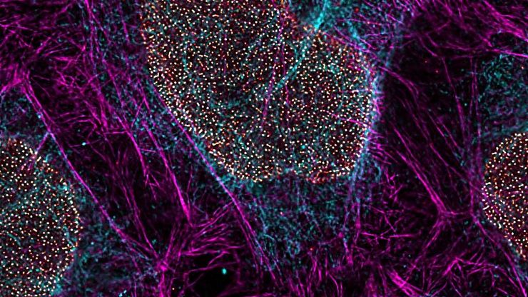
, microglia (TMEM119, IBA1), and Alzheimer’s-associated markers (β-amyloid and p-Tau217).")
利用大数据探索阿尔茨海默病的空间蛋白组
阿尔茨海默病是一种遗传性和散发性的神经退行性疾病,导致中晚年认知能力下降,特征为β-淀粉样蛋白斑块和 tau蛋白 缠结。由于治疗选择有限,新的研究策略至关重要。Cell DIVE 多重成像解决方案可以对阿尔茨海默病脑组织进行研究,揭示,可能新的研究方向。这里我们展示了 Cell DIVE 多重成像仪的图像查看器,用户能够直接在自己的浏览器中访问完整的阿尔茨海默病多重数据集。

利用大数据查看器揭示结肠癌隐藏的复杂性
结直肠癌是一种的重大健康负担。虽然手术初期有效,但部分患者会发展为预后不良的复发性继发疾病,需要采用免疫疗法等先进治疗手段。利用空间生物学方法,如 Cell DIVE 多重成像技术,可为开发新型治疗方案提供关键洞见。通过 Minerva 图像查看器在浏览器中访问完整的 Cell DIVE 数据集,进一步探索这些发现。

用大数据视角深入了解胰腺癌研究
胰腺癌由于其靠近主要器官难以分辨和难治疗,死亡率接近 40%,。这个研究探讨了胰腺导管腺癌(PDAC)的复杂生物学机制,研究了代谢、凋亡和免疫中肿瘤侵袭性的相关分子结构和空间决定因素。可以访问您的浏览器中的完整 Cell DIVE 数据集,以深入了解这些发现。

利用人工智能驱动的空间蛋白质组学绘制肿瘤免疫图谱
未经治疗肿瘤的空间图谱分析可呈现肿瘤免疫结构的整体特征,有助于理解治疗反应。具有免疫活性的小鼠模型对于识别肿瘤发生发展过程中免疫依赖性事件至关重要。要表征这些具有完整免疫系统及相互作用细胞组分的模型,需要采用多重标记分析技术。我们展示了一种基于人工智能的空间蛋白质组学方法,用于研究小鼠癌组织中的肿瘤-免疫互作机制。

阿尔茨海默病神经免疫相互作用的空间分析
阿尔茨海默病(AD)是一种复杂的神经退行性疾病,以神经原纤维缠结、β-淀粉样斑块和神经炎症为特征。这些功能障碍由局部免疫反应触发或加剧。因此,在空间背景下理解神经免疫相互作用对于阐明 AD 发病机制至关重要。本研究采用 Cell DIVE 多重成像技术和 Aivia 人工智能辅助空间分析工具,探究 AD 病理标志物周围免疫细胞的特征。

空间生物学指南
什么是空间生物学?在后组学时代,研究人员如何利用空间生物学工具来满足生物学问题日益增长的需求?本文简要概述了空间生物学及其技术,以及这一快速发展中的领域的关键研究问题。

使用空间多重化探测人类阿尔茨海默病皮层切片
阿尔茨海默病(AD)是最常见的神经退行性疾病,其特征是认知功能的逐渐下降。对 AD 大脑的空间分析可能揭示细胞关系,从而促进对疾病病因的更好理解。本研究捕捉了 AD 皮层组织成分的全球概述,并强调了 Cell DIVE 成像的简化工作流程,从数据采集到使用 Aivia 软件的基于人工智能的分析,最终实现更快的洞察。

通过开放多重化和细胞 DIVE 赋能空间生物学
空间生物学和多重成像工作流程在免疫肿瘤学研究中变得越来越重要。许多研究人员即使使用有效的工具和方案,也很难提高研究效率。我们将介绍研究人员如何利用开放式超多重免疫荧光的适应性,将 IBEX 成像与Cell DIVE 相结合,创造了一种名为 Cell DIVE-IBEX 的技术。它让这些研究人员能够调整现有的技术和试剂,并获得Cell DIVE 在其免疫肿瘤学研究中的可扩展性。
 tissue.")
基于人工智能的多重图像分析以探索结肠腺癌
在这项研究中,我们展示了一种利用Cell DIVE和AIVIA软件的空间生物学工作流程,以绘制结肠腺癌中的肿瘤免疫景观图。

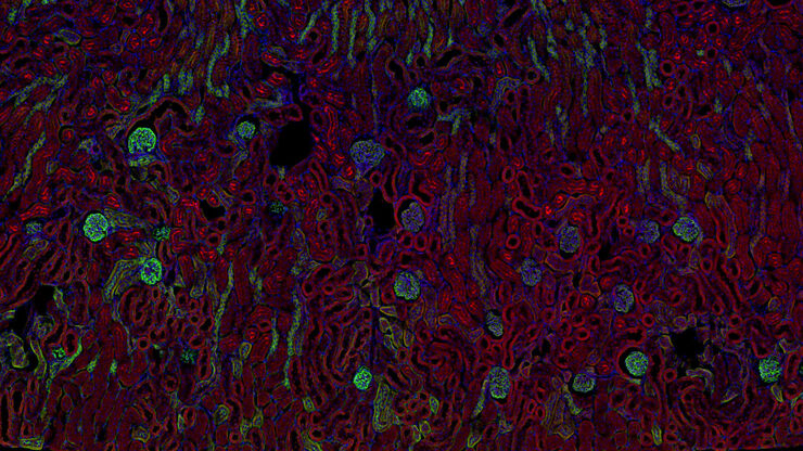
肿瘤空间微环境的元癌症分析
研究 TME中肿瘤、基质和免疫细胞之间的相互作用需要采用超多重免疫荧光成像方法。在这里,我们分析了一组Cell Signaling Technology(CST®)抗体,这些抗体针对肺癌、结肠癌和胰腺癌等癌症的标志物。通过Cell DIVE成像和Aivia中的聚类分析,我们确定了TME中的空间相互作用,包括组织特异性和共有的相互作用。
 tissue.")
通过成像和AI绘制结直肠癌的景观
结肠癌是一种高负担疾病。尽管进行了化疗干预和手术切除,但疾病可能会复发。了解结肠癌微环境对于改善治疗效果是必要的。在这里,我们使用空间生物学方法,通过Cell DIVE和 Aivia可视化结肠腺癌组织中的30个生物标志物。我们探讨了肿瘤组织的血管化、免疫细胞反应和细胞增殖。

IBEX、Cell DIVE 和 RNA-Seq:一种针对滤泡性淋巴瘤的多组学方法
在拉德特克等人最近的一项研究中,多组学空间生物学方法有助于揭示早期复发淋巴瘤患者的病情。
加速不同组织多重成像的发现
组织的多重成像对于肿瘤-免疫相互作用的研究以及人类细胞图谱等发现工作越来越重要。 欢迎加入我们的演讲,Andrea J. Radtke 博士解释了如何使用迭代漂白扩展多重性 (IBEX) 绘制组织图谱,并讨论了用于多重成像的广泛社区资源。

基于 AI 引导的多重二维数据向空间洞察的转化
Aivia 13 能够处理大型二维图像,使研究人员能够通过检测数百万个对象和自动聚类多达 30 个标记物,深入理解其表型周围的微环境。

利用蛋白质标记成像了解肿瘤异质性
Alison Cheung博士展示了如何利用蛋白质多重成像技术为癌症研究提供定量见解,与她一起探索肿瘤异质性和免疫细胞动态。

大脑的形状:阿尔茨海默病的空间生物学
阿尔茨海默病(AD)是一种神经退行性疾病,也是导致晚年认知障碍的常见原因。阿尔茨海默病的特征是出现含β-淀粉样蛋白的斑块和含磷酸化 tau 的神经纤维缠结。目前尚缺乏治疗和预防AD的有效疗法。我们将Cell DIVE与Cell Signaling Technology的抗体结合使用,检查了AD的突触过程并从空间上确定了神经胶质细胞和神经元等细胞,证明了超多标免疫荧光成像技术可用于探测AD大脑。

探索多重生物成像如何推进癌症研究
观看行业和学术专家进行的内容丰富的讨论,分享他们在研究中使用多重成像技术的知识。了解多重成像技术如何通过发现以前难以捉摸的分子洞察力,彻底改变肿瘤学、神经学和免疫学。利用先进的成像技术深入了解组织微环境,从而对代谢紊乱和癌症等疾病有新的认识。

空间生物学:理解全景
空间生物学:了解分子、细胞和组织在原生空间环境中的组织和相互作用

借助多重成像深入了解胰腺癌的复杂性
胰腺癌是一种很难治疗的肿瘤疾病。Cell DIVE多重成像可以视觉呈现30种生物标志物以探测胰管癌的微环境。此面板可以检查肿瘤组织多个层级的问题,包括淋巴细胞、血管新生、转移、侵袭、炎症、缺氧、代谢和免疫。多重成像和分析可以对肿瘤组织中的许多生物活动提供更为深入的洞察信息,从而可以深入研究这些信息。

化繁为简:多重成像中的抗体
了解抗体对于多重成像研究的重要意义,以及如何规划并建立自己的抗体组合

多标成像的类型、优势和应用
与传统显微镜相比,多标成像技术可以观察到更多的生物标记物,是一种从人体组织样本中提取信息的新兴且令人兴奋的方法。通过同时观察许多生物标记物,可以协同探索以前只能单独探索的生物通路,并识别和探究复杂的组织和细胞表型。目前已有许多不同的多标成像方法,每种方法都采用不同的方法来实现更高的染色。

Cell DIVE已验证的抗体将使您对实验结果产生信心
Cell DIVE超多标组织成像分析整体解决方案包括经严格验证的350+抗体资源库,高灵敏度高特异性的应用于Cell DIVE循环染色成像中。抗体验证的方法可以帮助您找到合适的抗体以及最佳的实验条件,快速的让您开展超多标成像分析的实验。抗体库中的每种抗体都经过严格的三步验证过程,(a)评估在FFPE上的表现性能;(b)确定其最佳的染色条件以及是否可用作直标抗体;(c)探究由于Cell…
癌症研究
癌症是一种复杂的异质性疾病,由于细胞生长失控而引起。 一个或一组细胞的基因和表观遗传的变化破坏了正常功能,导致细胞自发、不受控制地生长和增殖。

高级组织成像& 分析
利用徕卡显微系统的先进成像解决方案,深入了解组织结构和功能,从而加深对空间生物学和疾病机制的理解。
生物制药
对于生物制药行业,Leica 解决方案有助于加快药物发现,增强细胞分析,并支持符合法规的数据完整性。
细胞分析
先进的显微成像解决方案和用于细胞分析的人工智能分析软件有助于研究人员深入了解细胞功能和亚细胞结构。
应用领域
细胞分析
先进的显微成像解决方案和用于细胞分析的人工智能分析软件有助于研究人员深入了解细胞功能和亚细胞结构。
生物制药
对于生物制药行业,Leica 解决方案有助于加快药物发现,增强细胞分析,并支持符合法规的数据完整性。
高级组织成像& 分析
利用徕卡显微系统的先进成像解决方案,深入了解组织结构和功能,从而加深对空间生物学和疾病机制的理解。
