艺术保护&文化遗产

利用先进的成像解决方案,分析、修复、保护与记载艺术作品或文物,需要复杂的技术技能和顶级成像解决方案,尤其是以无接触、非破坏性的方式。结合数码相机技术和图像分析软件,徕卡显微系统的艺术保护显微镜解决方案专为不同领域的修复学家、艺术历史学家、考古学家以及保护工作室和博物馆的专家所设计。所有的显微镜解决方案都对详细准确地分析样品、显微分析结构及材料并存档进行了优化。

欢迎联系我们

如果您对徕卡显微系统绘画和艺术品保护显微镜感兴趣,需要专家建议,欢迎联系我们。

使您不会错过任何细节

徕卡显微系统专为不同领域的修复学家、艺术历史学家、考古学家以及相关领域的专家提供各种解决方案。对详细准确地分析样品、显微分析结构及材料并存档,全部进行了优化。

为专家提供专业知识

显微镜解决方案尤其适用于特殊要求,与徕卡显微系统的数码相机技术和图像分析软件相结合,成为保护工作室和博物馆的许多专家常用的专业工具。

为了更好地了解每个应用领域不同的方法和技术,我们列出了下列产品,帮助您进行宝贵的艺术珍品以及不可替代的原物方面的工作。

颜料分析

检查油漆层和油漆颜料时,需要明亮冷照明的高分辨率、高性能显微镜。徕卡显微系统提供功能强大的显微镜解决方案,荧光照明选项和易于使用的分析软件相结合,用于优化检测、分析和存档。

X-radiograph (detail) with the revealed head of a woman painted over.

绘画

详细精确的清洁和修复画作,包括使用为各种绘画类型和尺寸定制的精确数码和立体显微镜。用于绘画清洁和修复的徕卡显微镜提供可变变焦位置,使您可以 即使用低放大倍率,也能分析微小的细节。大型绘画可选用落地式配件。

材料

矿物/化石

矿物和化石保护需要通过高性能的立体显微镜和研究显微镜,进行精细的图像处理和分析。无论您需要三维观测微结构还是需要分析薄切片,徕卡显微系统都能提供满足您实际需要的多功能显微解决方案。

纸张/书籍/照片

利用徕卡的一系列显微镜解决方案,揭示和分析印刷和整理技术、手稿或修饰的独特细节,并以三维方式查看最精细的纸张结构。这些创新的数字和立体显微镜结合了最高的分辨率和最大的景深,以支持您研究和修复这些珍贵的遗产项目所需的获取、存储、注释和记录。

玻璃/陶瓷/石材

当您检查、清除或重建玻璃、陶瓷或石头时,您需要定制的、高分辨率的显微镜解决方案,使您能够分析不同尺寸和形状的物体。徕卡显微系统公司提供各种立体和研究型显微镜,并根据您的需要提供不同的照明方法和便携式支架系统。

金属

通过徕卡显微系统的定制化、经济实惠的系统解决方案,可以检查金属物品的腐蚀迹象、污垢沉积、加工或处理技术,或者分析金属结构,确定合金成分。独具特色的立体显微镜和研究向显微镜与数码相机以及易于使用的三维软件相结合,支持您对金属物品的分析、清洁和记录。

木材

利用徕卡立体显微镜和研究显微镜解决方案,清洁和修复油漆或清漆的木制物品,并确定应用层的结构和零件。这些灵活的解决方案和台面或落地支架、数码相机以及易于使用的分析软件相结合,为您的木材保护工作提供支持。

银色烛台

用于修复和保护金属物品,例如这个来自十九世纪的银烛台。利用显微镜分析受损区域、清洁表面并存档。在显微镜下,银烛台的锈蚀区域中发现了一个人造的铜锈。

©阿姆斯特丹国家博物馆

Rock crystal (quartz) and rutile needles enclosed in rock crystal (quartz), from Minas Gerais, Brazil. Because of the light metallic lustre of the rutile needles (titanium oxide, TiO2), the trade name of this quartz variety is “platinum quartz”. The picture shows a first generation rock crystal inclusion that was enclosed by a second generation of quartz. Although the inclusion and the host material have the same refractive index, the enclosed quartz is easily recognizable due to a thin film of air at the interfaces. Width of image: approx. 6 mm, transmitted light, crossed polarizers, first-order red compensator. © Michael Hügi

岩晶

来自于巴西米纳斯吉拉斯州的水晶(石英)和包含在水晶(石英)中的Anchor金红石针。由于金红石针(二氧化钛TiO2)上明亮的金属光泽,因此这种石英的商品名称为“铂石英”。该图表示被包围在第二代石英中的第一代水晶杂质。虽然杂质和主体材料具有相同的折射率,但是由于被包围的石英在交界面上有一层空气薄膜,因此可以很容易地辨认出。图像宽度:约为6mm,透射光,正交偏光,一阶红色补偿。

© Michael Hügi

For restoration of ancient photos, high-end stereomicroscopes combined with digital cameras are used for acquiring, storing, annotating and displaying high quality images of our heritage which needs restoration. The microscopic analysis supports the recognition of the original material, the analysis of the conservation status and effects of different methods of restoration. © Alinari, Florence

古代照片

为了修复老照片,可采用高端立体显微镜结合数码相机,用于采集、存储、注释和展示保留下来需要修复的高品质图像。显微分析支持识别原材料,分析保护现状和不同修复方法的修复效果。

© Alinari, Florence

Jacob Jordaens (1593–1678), “The Tribute Money. Peter Finding the Silver Coin in the Mouth of the Fish”, also called “The Ferry Boat to Antwerp” (Oil on canvas, 279.5 by 467 cm. With a stereomicroscope combined with a floor stand the paint surface has been investigated and analyzed. © Statens Museum for Kunst, Copenhagen

“献金.彼得在鱼嘴中发现了银币”

Jacob Jordaens (1593–1678),“献金.彼得在鱼嘴中发现了银币”,也称为“到安特卫普的渡船”(布面油画,279.5×467cm)。利用带有落地支架的立体显微镜,研究并分析油画表面。

© 丹麦国家美术馆,哥本哈根

Willem van Honthorst (1594-1666) “Portrait of a Young Man Holding a Plumed Hat in his Right Hand” The almost life-size portrait dating from the first half of the 17th century requires extensive restoration. The microscope examination shows the rough granular surface of this Dutch painting which is a result of a later application of varnish with poorly dissolved components. ©Sammlungen des Fürsten von und zu Liechtenstein, Vaduz–Wien

“右手持一顶羽毛帽的年轻人的肖像画”

Willem van Honthorst (1594-1666) “右手持一顶羽毛帽的年轻人的肖像画” 始于第17世纪上半叶的几乎和真人一样大小的肖像画,需要大量修复。使用了清漆与难以溶解的成分之后,显微镜检查可以展示出这种荷兰绘画的粗糙的颗粒状表面。

© Sammlungen des Fürsten von und zu Liechtenstein, Vaduz–Wien

Rock crystal (quartz) and rutile needles enclosed in rock crystal (quartz), from Minas Gerais, Brazil. Because of the light metallic lustre of the rutile needles (titanium oxide, TiO2), the trade name of this quartz variety is “platinum quartz”. The picture shows a first generation rock crystal inclusion that was enclosed by a second generation of quartz. Although the inclusion and the host material have the same refractive index, the enclosed quartz is easily recognizable due to a thin film of air at the interfaces. Width of image: approx. 6 mm, transmitted light, crossed polarizers, first-order red compensator. © Michael Hügi
For restoration of ancient photos, high-end stereomicroscopes combined with digital cameras are used for acquiring, storing, annotating and displaying high quality images of our heritage which needs restoration. The microscopic analysis supports the recognition of the original material, the analysis of the conservation status and effects of different methods of restoration. © Alinari, Florence
Jacob Jordaens (1593–1678), “The Tribute Money. Peter Finding the Silver Coin in the Mouth of the Fish”, also called “The Ferry Boat to Antwerp” (Oil on canvas, 279.5 by 467 cm. With a stereomicroscope combined with a floor stand the paint surface has been investigated and analyzed. © Statens Museum for Kunst, Copenhagen
Willem van Honthorst (1594-1666) “Portrait of a Young Man Holding a Plumed Hat in his Right Hand” The almost life-size portrait dating from the first half of the 17th century requires extensive restoration. The microscope examination shows the rough granular surface of this Dutch painting which is a result of a later application of varnish with poorly dissolved components. ©Sammlungen des Fürsten von und zu Liechtenstein, Vaduz–Wien

相关文章

艺术保护&文化遗产

作为徕卡显微系统有限公司 的信息门户,ScienceLab (徕卡课堂) 提供许多关于显微技术主题的科研和教学材料。其内容宗旨在于为初学者、有经验的从业者和科学家等的日常工作和实验提供支持。

更多文章
带有大量夹杂物的原生刚玉玻璃图像。这是使用偏振对比显微镜拍摄的。

利用偏光显微镜优势确保玻璃质量

玻璃是已知最古老的材料之一。如今,玻璃被广泛应用于各种领域,如光学仪器、门窗、太阳能电池板、食品、饮料和药品容器等,因此必须符合严格的玻璃质量标准,尤其是光学玻璃。利用偏光显微镜对平板玻璃、中空玻璃和压制玻璃进行质量控制既快捷又经济。无需进行耗时的样品制备,即可对结节、金属、晶体夹杂物和气泡等缺陷进行分析。
Some 2D measurements, e.g., lengths and areas, made on a PCB sample with a Leica measurement microscope using the Enersight software.

如何选择合适的测量显微镜

使用测量显微镜,用户可以测量样品特征的二维和三维尺寸,这对检测、质量控制、故障分析和研发&D 至关重要。然而,选择合适的显微镜需要评估应用需求以及显微镜的性能、易用性和灵活性。 如今,测量通常以数字方式进行,即使用带有摄像头和软件的显微镜,图像显示在显示器上,而不是通过目镜网线,从而提高了精度和可重复性。使用合适的测量显微镜可靠、快速地分析样品。
Example of calibrating a microscope at a higher magnification value using a stage micrometer.

显微镜测量校准:为什么要这样做?

显微镜校准可确保检测、质量控制 (QC)、故障分析和研发 (R&D) 所需的测量准确一致。本文介绍了校准步骤。使用参照物进行校准可使结果具有可重复性,并有助于确保与准则和标准一致。为了获得准确一致的结果,建议校准显微镜并定期检查。如有需要,可向校准专家寻求支持。
Optical microscope image, which is a composition of both brightfield and fluorescence illumination, showing organic contamination on a wafer surface. The inset images in the upper left corner show the brightfield image (above) and fluorescence image (below with dark background).

晶圆表面光刻胶残留与有机污染物可视化检测

随着半导体集成电路 (IC) 的尺寸缩小到 10 纳米以下,在晶圆检测过程中有效检测光刻胶残留物等有机污染物和缺陷变得越来越重要。光学显微镜仍是常用的检测方法,但对于有机污染而言,明视野和其他类型的照明都有其局限性。本文讨论了在半导体行业的质量控制、故障分析和研发&D 过程中,如何利用荧光显微镜有效检测晶片上的光刻胶残留物和其他有机污染物。
Image of magnetic steel taken with a 100x objective using Kerr microscopy. The magnetic domains in the grains appear in the image with lighter and darker patterns. A few domains are marked with red arrows. Courtesy of Florian Lang-Melzian, Robert Bosch GmbH, Germany.

Rapidly Visualizing Magnetic Domains in Steel with Kerr Microscopy

The rotation of polarized light after interaction with magnetic domains in a material, known as the Kerr effect, enables the investigation of magnetized samples with Kerr microscopy. It allows rapid…
Region of a patterned wafer inspected using optical microscopy and automated and reproducible DIC (differential interference contrast). With DIC users are able to visualize small height differences on the wafer surface more easily.

6 英寸晶片检测显微镜,可靠的观察微小高度差

本文介绍了一种 6 英寸晶圆检测显微镜,无论用户的技术水平如何,它都能自动进行可重复的 DIC(微分干涉对比)成像。集成电路 (IC) 芯片和半导体元件的制造需要晶圆检测,以确保不存在影响性能的缺陷。通常使用光学显微镜进行质量控制、故障分析和 R&D 检测。为了有效地观察晶圆上结构之间的微小高度差,可以使用 DIC。
Automated wafer loader using carbon fiber end-effectors for safer handling.

无需用手接触即可安全装载晶片,进行显微镜检测

本文介绍了用于显微镜检测的自动硅晶片装载如何帮助改进微电子工艺控制和生产效率。人工搬运晶圆很可能会损坏脆弱的晶圆表面,从而增加成本,而自动化搬运则能确保更安全、更具成本效益的生产。自动晶片装载机在显微镜检测和制造方面的优势概述如下。
Image of burrs (red arrows) 
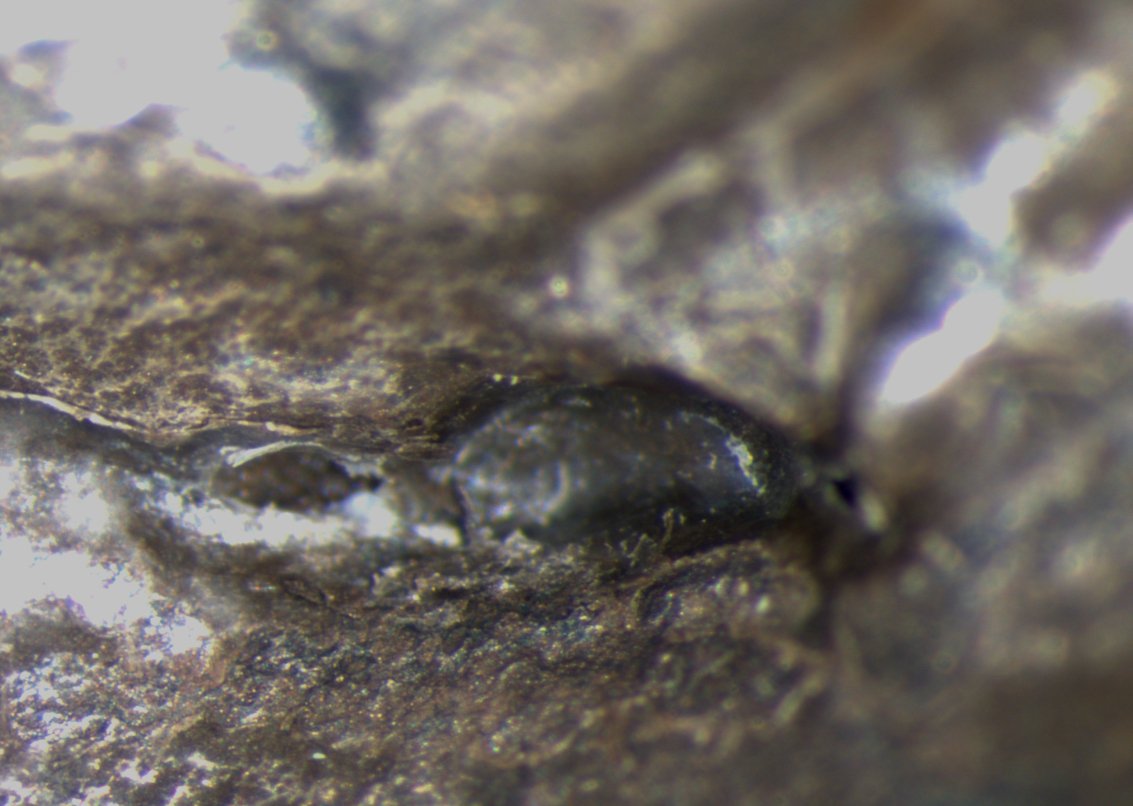
at the edge of a battery electrode acquired with a DVM6 digital microscope.

电池制造过程中的毛刺检测

毛刺是电池电极片边缘可能出现的缺陷,例如在制造过程中的分切环节。它们可能会因诸如短路等故障导致电池性能下降,并引发安全和可靠性问题。毛刺检测是电池生产质量控制的重要部分,对于生产具有可靠性能和寿命的电池至关重要。通过适当照明的光学显微镜可以在生产过程的关键步骤中快速可靠地对电极上的毛刺进行视觉检测。
Quality assurance during production in a manufacturing plant.

跨行业的质量保证改进

精确是最重要的。试想一下,心脏起搏器在运行过程中发生故障,或者半导体缺陷导致关键系统崩溃。在医疗设备、电子产品和半导体等行业,误差几乎为零。质量保证(QA)不再仅仅是一项监管要求,而是一项推动业务成功和保护品牌完整性的战略优势。
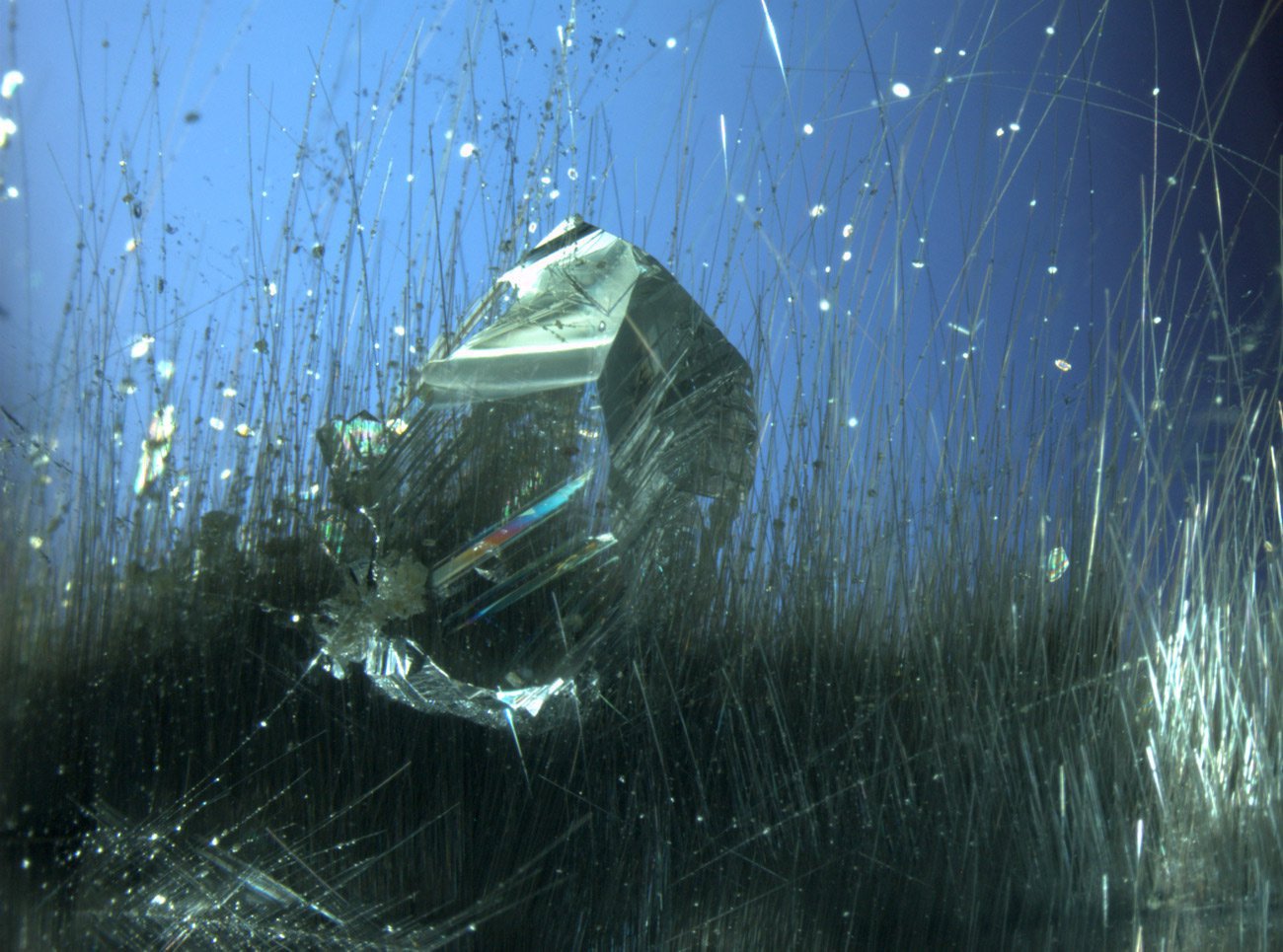
用 Leica 显微镜使用圆偏振光对径向生长的糖晶体进行成像。

偏振光显微镜影像图集

偏振光显微镜(又称为偏光显微镜)是一种应用于不同领域的重要方法,包括研究和质量保证。它不仅仅是在高倍率和高分辨率下产生图像,这通常是用普通光学显微镜完成的。 通过检查样本的形状、结构、颜色、双折射和进一步的光学性质,可以获得有关样本结构、光学性质和成分的附加信息。
Scroll to top