
Filter articles
标签
Loading...
.")
利用光片显微技术聚焦三维长时程成像
长时程三维成像揭示了复杂的多细胞系统是如何生长和发育的,以及细胞是如何随着时间的推移而移动和相互作用的,从而揭示了发育、疾病和再生方面的重要知识。光片显微镜一次只照射样品的一个薄片,大大减少了光损伤,保护了样品的活性。这种温和的高速技术可在数小时甚至数天内提供清晰的体数据,使研究人员能够实时捕捉生物学的发展过程。
Loading...

生理学图片库
生理学是关于生物体内的过程和功能。生理学研究的重点是生物体器官、组织或细胞的活动和功能,包括所涉及的物理和化学现象。在此,我们以不同的样本为例,向您展示与生理学有关的图片。
Loading...
组织图片库
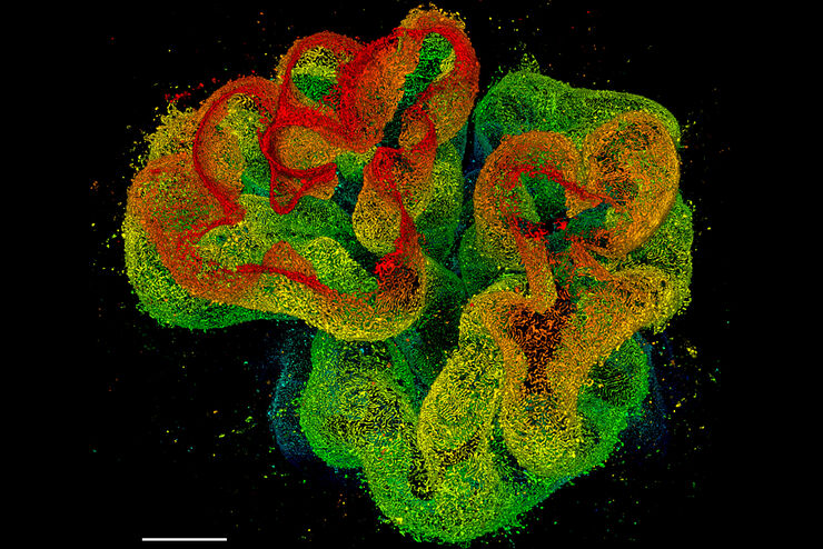
对动物和人体组织进行视觉分析对于了解癌症或神经变性等复杂疾病至关重要。从基本的免疫组化到体内成像,共聚焦显微镜和先进的模式可以让人们了解细胞、生物分子及其在环境中的相互作用。
 and astrocytes (green) in a cortical spheroid derived from human induced pluripotent stem cells.")

Loading...

Image Gallery: THUNDER Imager
To help you answer important scientific questions, THUNDER Imagers eliminate the out-of-focus blur that clouds the view of thick samples when using camera-based fluorescence microscopes. They achieve…
Loading...
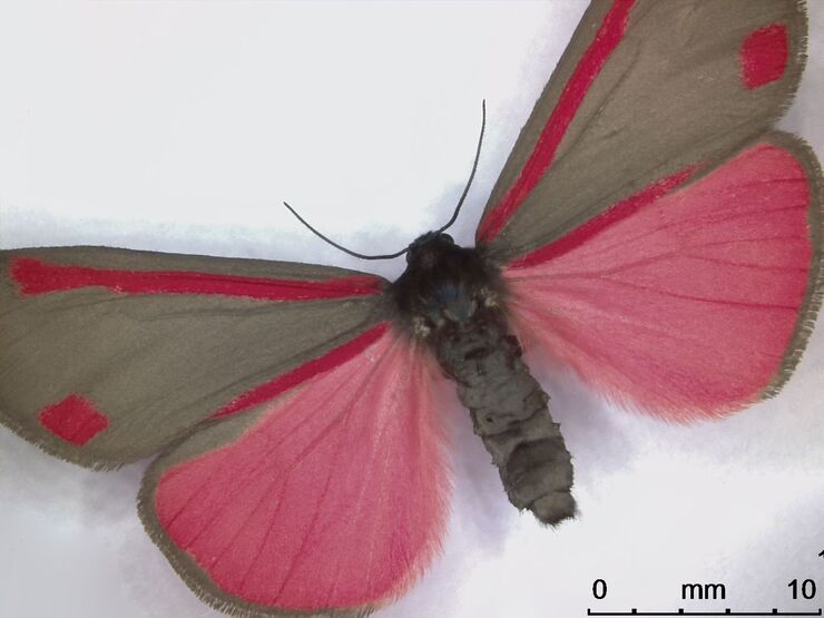
Image gallery: Life Science Imaging with the Leica DVM6 Digital Microscope
Digital microscopes can be a great help in life science applications such as the documentation in botany, entomology studies and crop science, or the digitization of museum collections. The image…
