工业

工业

工业

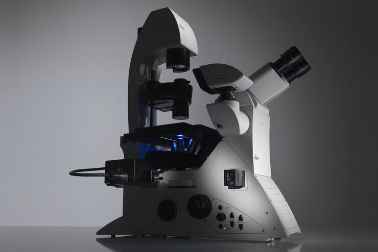
深入探讨有关工业和病理学领域的高效检测、优化工作流程和提高人体工学舒适度的文章和网络研讨会。涉及的主题包括质量控制、材料分析、病理学显微镜等。在这里您可以获得有关使用前沿技术提高生产力和优化质量以及准确地进行病理学诊断的干货。
These images illustrate the need for multiple z-slices to capture all gH2Ax foci in a given cell and get an accurate count.

Development and Derisking of CRISPR Therapies for Rare Diseases

This on-demand presentation by Dr. Fyodor Urnov and Dr. Sadik Kassim, originally delivered at ASGCT 2025, focused on a critical challenge in genetic medicine: how to scale CRISPR therapies from…
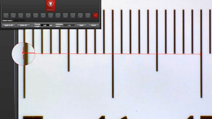
Example of calibrating a microscope at a higher magnification value using a stage micrometer.

Microscope Calibration for Measurements: Why and How You Should Do It

Microscope calibration ensures accurate and consistent measurements for inspection, quality control (QC), failure analysis, and research and development (R&D). Calibration steps are described in this…
Mouse brain slice which was immunostained with GFAP-A647 and imaged using a THUNDER Imager Tissue. Courtesy of H. Xu, University of Pennsylvania, Philadelphia, USA.

神经科学研究解决方案

您的工作是更好地了解神经退行性疾病,还是研究神经系统的功能? 了解如何使用徕卡显微系统的成像解决方案取得突破。

斑马鱼研究

为了在筛选、分拣、操作和成像过程中获取高质量结果,您需要观察细节和结构,从而为您的下一步研究做出正确的决策。 徕卡体视显微镜和透射光底座以出众的光学器件和优良的分辨率而闻名,是全世界研究学者的首选。
Zebrafish-embryo image captured using a THUNDER Imager Tissue and live instant computational clearing.

Improving Zebrafish-Embryo Screening with Fast, High-Contrast Imaging

Discover from this article how screening of transgenic zebrafish embryos is boosted with high-speed, high-contrast imaging using the DM6 B microscope, ensuring accurate targeting for developmental…
Transfection using the Uncommon Bio reprogramming system. Image acquired using the THUNDER Imager 3D Cell Culture with THUNDER Large Volume Computational Clearing (LVCC) applied. Image courtesy of Samuel East, Uncommon Bio.

利用新型可扩展的干细胞培养设计未来

具有远见卓识的生物技术初创企业 Uncommon Bio 正在应对世界上最大的健康挑战之一:食品可持续性。在这次网络研讨会上,干细胞科学家塞缪尔-伊斯特(Samuel East)将展示他们如何使细胞农业的干细胞培养基既安全又经济可行。了解他们如何将培养基成本降低 1000 倍,并开发出不含动物成分、食品安全的 iPSC 培养基。
Area of a printed circuit board (PCB) which was imaged with extended depth of field (EDOF) using digital microscopy.

如何形成清晰的图像

在显微镜检查中,景深常被看做经验参数。在实际操作中,会根据数值孔径、分辨率和放大率之间的相关性确定该参数。为了获得最佳视觉效果,现代显微镜的调节设备在景深和分辨率之间实现了最佳平衡,这两个参数在理论上呈负相关。
Stripe assay performed on a THUNDER Imager Cell. Courtesy of Maria Carrasquero Ordaz, University of Oxford.

揭示神经元迁移的分子奥秘

研究发育中大脑神经元向生态位迁移可采用多种方法。在本场研讨会中,牛津大学的专家们将展示他们用于阐明神经发育期间神经元向皮层功能层迁移的分子机制的显微技术与实验方法。理解这些过程将有助于更深入地认识健康大脑的发育机制,并可能为神经发育障碍提供更优治疗方案。
Image of magnetic steel taken with a 100x objective using Kerr microscopy. The magnetic domains in the grains appear in the image with lighter and darker patterns. A few domains are marked with red arrows. Courtesy of Florian Lang-Melzian, Robert Bosch GmbH, Germany.

使用克尔显微镜快速可视化钢中的磁畴

磁性材料中磁畴与偏振光相互作用后光的旋转,称为克尔效应,使得使用克尔显微镜对磁化样品进行研究成为可能。它可以快速可视化材料表面的磁域。对于用于电气和电子设备的磁性材料(例如钢合金)的高效研发和质量控制,克尔显微镜可以发挥重要作用。本文详细描述了如何使用克尔显微镜对钢合金晶粒中的磁域进行成像。
Scroll to top