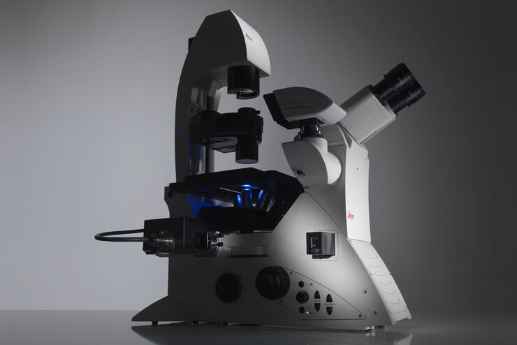
THUNDER Imager Tissue全景组织显微成像系统
THUNDER Imaging Systems
产品
首页
Leica Microsystems
THUNDER Imager Tissue全景组织显微成像系统
实时解构3D生物微观世界*
阅读我们的最新文章
斑马鱼研究
为了在筛选、分拣、操作和成像过程中获取高质量结果,您需要观察细节和结构,从而为您的下一步研究做出正确的决策。
徕卡体视显微镜和透射光底座以出众的光学器件和优良的分辨率而闻名,是全世界研究学者的首选。

Improving Zebrafish-Embryo Screening with Fast, High-Contrast Imaging
Discover from this article how screening of transgenic zebrafish embryos is boosted with high-speed, high-contrast imaging using the DM6 B microscope, ensuring accurate targeting for developmental…
 acquired using THUNDER Imager Live Cell. Image courtesy of Janina Kaspar and Irene Santisteban, Schäfer Lab, TUM.")
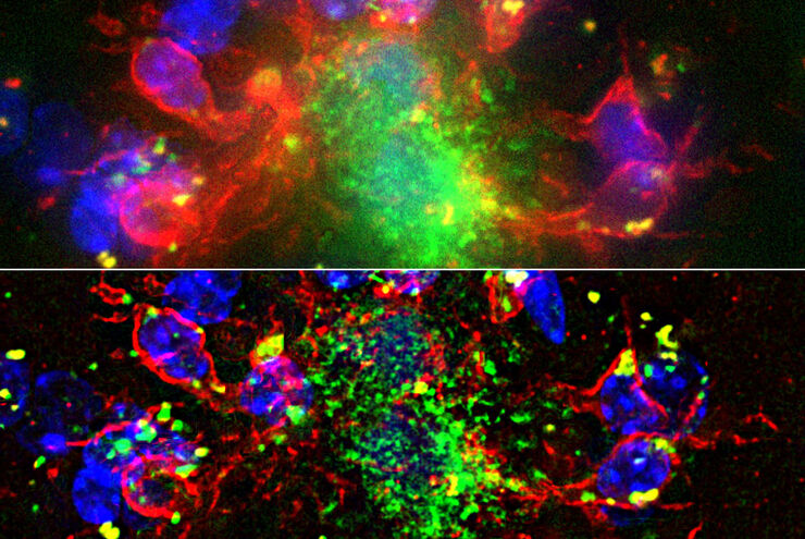
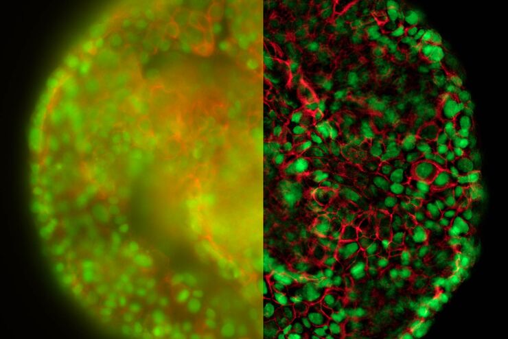
研究大脑健康的成像类器官模型
小胶质细胞是特化的脑驻留免疫细胞,在大脑发育、平衡和疾病中发挥着至关重要的作用。然而,到目前为止,模拟人脑环境与小胶质细胞之间相互作用的能力还非常有限。
. Image courtesy of Prof. Hui Guo, School of Life Sciences, Central South University, China")
显微镜如何帮助研究机械感受和突触通路
Tobi Langenhan教授使用显微镜研究突触蛋白质组合体,研究粘附性GPCR的机械感受特性,并了解蛋白质动力学及其空间相互作用。

神经科学显微镜面临哪些挑战?
显微镜是神经科学研究领域的强大工具。不过,当涉及到对神经过程进行成像以及使用不同的样品类型(例如厚神经组织或脑类器官)时,科研人员可能会面临到很多挑战。这本30页的电子书包含众多真实的案例,以讨论我们最常见到的一些挑战,同时展示了如何使用THUNDER 成像技术克服这些挑战。

超越反卷积
宽场荧光显微镜通常用于视觉呈现生命科学样本中的结构并获取重要信息。利用荧光蛋白或染料,以高度特异性的方式标记离散的样本部分。为了充分了解某种结构,可能需要以三维方式呈现,但这会对使用显微镜带来某些挑战。

从概览中查找相关样本细节
在从图像到图像的搜索中切换到快速查看整个样本概览,并即刻识别重要的样本细节。利用这些知识,使用载玻片、培养皿和多孔板的模板自动设置高分辨率图像采集。LAS X Navigator软件像是样本细胞的GPS,总能为用户指明通向高质量数据的清晰路径,这是生命科学平台STELLARIS和THUNDER成像仪上的一款强大的导航工具。LAS X Navigator支持将宽场、立体或共聚焦实验与舞台应用相结合。

精确分析宽视野荧光图像
利用荧光显微镜的特异性,即便是使用厚样品和大尺寸样品,研究人员也能够快速轻松地准确观察和分析生物学过程和结构。然而,离焦荧光会提高背景荧光,降低对比度,影响图像的精确分割。THUNDER 与Aivia 的组合可以有效解决这一问题。前者可以消除图像模糊,后者会使用人工智能技术自动分析宽视野图像,提高操作速度和精确性。下面,我们来详细了解下这一协作方法。

High-resolution 3D Imaging to Investigate Tissue Ageing
Award-winning researcher Dr. Anjali Kusumbe demonstrates age-related changes in vascular microenvironments through single-cell resolution 3D imaging of young and aged organs.

优化 THUNDER 平台以实现高内涵玻片扫描
随着对全组织成像需求的不断增长以及对不同生物标本中 FL 信号定量的需要,HC 成像技术的极限受到了考验,而核心设备的用户可培训性和易用性则成为了成本和效率的问题。在这里,我们展示了在我们的设施中为THUNDER平台开发的可行工作流程,以支持从 KO-小鼠组织分析到人类癌症的各种研究环境需求。

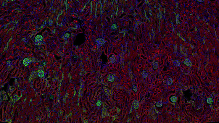
生理学图片库
生理学是关于生物体内的过程和功能。生理学研究的重点是生物体器官、组织或细胞的活动和功能,包括所涉及的物理和化学现象。在此,我们以不同的样本为例,向您展示与生理学有关的图片。
暗场显微镜
此外,在对材料样本进行成像时,暗场显微镜还能增强图像对比度。暗场光学对比法利用生物标本结构或材料样本的不均匀特征产生的光散射或衍射。

发育生物学图片库
发育生物学探索复杂生物体从胚胎到成年的发育过程,以详细了解疾病的起源。图库的这一类别显示有关发育生物学的图像,即通常以昆虫、蠕虫、动物和植物为研究对象的图像。

自适应反卷积与 Computational Clearing 结合的力量
反卷积是一种计算方法,用于恢复被点扩散函数(PSF)和噪声源破坏的物体图像。在本技术简介中,您将了解徕卡显微系统提供的反卷积算法如何帮助您克服宽视场 (WF) 荧光显微镜中由于光的波动性和光学元件对光的衍射而造成的图像分辨率和对比度损失。探索由用户控制或自动反卷积的方法,查看并解析更多的结构细节。
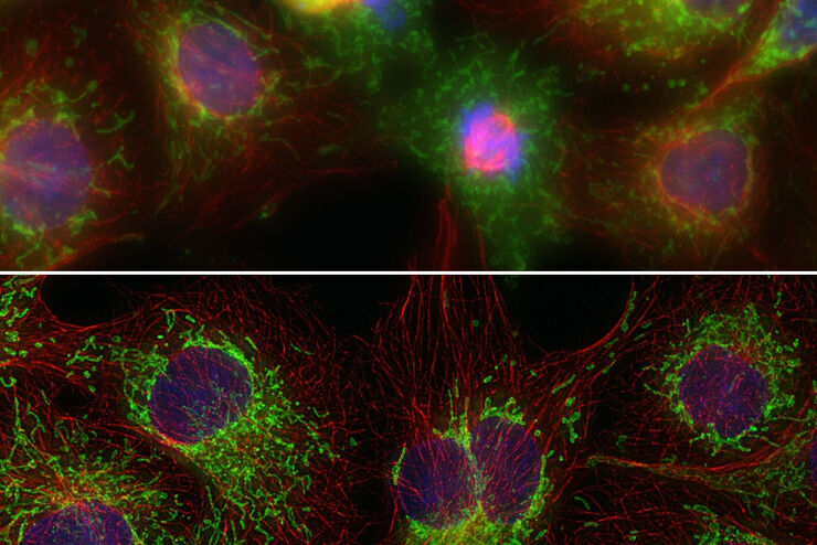
改进成像技术以了解细胞器膜细胞动态
了解正常组织和肿瘤组织中的细胞功能,是推动潜在治疗策略研究和了解某些治疗失败原因的关键因素。单细胞分析在生物医学研究中至关重要,它能揭示在癌症等复杂疾病中哪些细胞和分子通路发生了改变。

Image Gallery: THUNDER Imager
To help you answer important scientific questions, THUNDER Imagers eliminate the out-of-focus blur that clouds the view of thick samples when using camera-based fluorescence microscopes. They achieve…

从器官到组织再到细胞:使用宽场显微镜分析 3D 标本
在传统的宽场显微镜下从厚的三维样本中获取高质量的数据和图像是具有挑战性的,因为存在失焦光的干扰。在本次网络研讨会中,Falco Krüger 展示了THUNDER成像仪如何通过Computational Clearing技术使这一切成为可能。
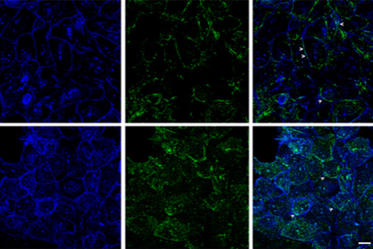
:内皮细胞,IsoB4(红色):血管,小胶质细胞抗 GFAP 抗体(蓝色):星形胶质细胞。样本由美国南旧金山 Genentech 公司的 Jeremy Burton")
消翳现真—突破传统宽场成像的极限
许多软件包都包含成像优化算法,通过降低背景噪声来增强图像特征的对比度。从 WF 图像中去除背景噪声最常用的方法是滚动球和滑动抛物面。近期徕卡显微系统公司推出了其自主研发的成像优化技术—即时成像解析(ICC),该技术已集成于所有徕卡THUNDER宽场成像平台。
Factors to Consider When Selecting a Research Microscope
An optical microscope is often one of the central devices in a life-science research lab. It can be used for various applications which shed light on many scientific questions. Thereby the…
Factors to Consider When Selecting a Research Microscope
An optical microscope is often one of the central devices in a life-science research lab. It can be used for various applications which shed light on many scientific questions. Thereby the…
Computational Clearing - 增强3D标本成像
本次网络研讨会旨在阐明有助于THUNDER显微成像仪实现三维样品可视化的关键规格,并改进研究人员的成像相关工作流程。
癌症研究
癌症是一种复杂的异质性疾病,由于细胞生长失控而引起。 一个或一组细胞的基因和表观遗传的变化破坏了正常功能,导致细胞自发、不受控制地生长和增殖。
THUNDER成像:高效、灵活、易操作,让您的日常成像工作流更轻松
本次网络研讨会将展示 THUNDER 在许多不同生命科学应用中的多功能性和性能:从计数视网膜切片中的细胞核和癌组织切片中的 RNA 分子,到监测阿拉伯芥幼苗中的钙波等等。
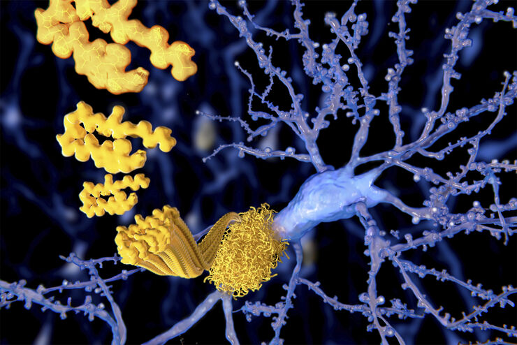
阿尔茨海默斑块:厚切片中的快速可视化
超过 60%的所有诊断为痴呆症的病例归因于阿尔茨海默病。该疾病的典型特征是脑组织的组织学改变。目前尚无治愈该疾病的方法。一些治疗方法试图减缓致命的进程或缓解患者的症状。梅赫达德·沙姆鲁博士的实验室研究病理性脑功能,旨在为阿尔茨海默病的新疗法的发现做出贡献。他们使用这种疾病的小鼠模型研究炎症在阿尔茨海默病进展中的作用。这需要对厚的未清除脑组织进行成像。
清晰对比、无雾的 3D 样本实时图像
历史上,宽场显微镜并不适合对大样本/标本体积进行成像。图像背景(BG)主要来源于观察样本的失焦区域,显著降低了成像系统的对比度、有效动态范围和最大可能的信噪比(SNR)。记录的图像显示出典型的雾霭,并且在许多情况下,无法提供进一步分析所需的细节水平。处理厚三维样本的研究人员要么使用替代显微镜方法,要么尝试通过后处理一系列图像来减少雾霭。
