Filter articles
标签
产品
Loading...

用于三维生物成像的集成连续切片与冷冻电镜工作流程
本场网络研讨会探讨了集成化工具如何支持从样品制备到图像分析的电子显微镜全流程。专家Andreia Pinto博士、Adrian Boey博士与Hoyin Lai博士将介绍UC Enuity超薄切片机和Aivia图像分析平台,并演示这些工具如何同时适用于常温与低温实验环境。会议内容包含阵列断层成像、基于深度学习的图像分割、以及生物成像中cryo-lift-out工作流程的实际案例解析。
Loading...

通过Cryo-EM(冷冻电镜)和 CryoFIB(冷冻聚焦离子束) 揭示钠电池退化机制
探索低温电镜和聚焦离子束技术如何揭示钠电池界面的内在结构。本次研讨会将提出基于隔膜渗透(而非枝晶生长)的新型退化模型,并解析电解液溶剂如何影响界面稳定性与电池性能。
Loading...

从显微镜到电镜:完整的冷冻光电联用工作流程
在题为“多模态玻璃化征程,从实验台到电子显微镜的冷冻关联工作流程”的网络研讨会上,专家团队(Edoardo D'Imprima、Zhengyi Yang、Andreia Pinto 和 Martin…
Loading...

如何成功应用Coral life
许多电子显微镜(EM)工作流程始于样品固定,随后进行样品准备和电镜成像。然而,表现出有趣行为的样品往往很罕见,找到“合适的细胞”可能耗时且繁琐。活细胞光电联用工作流程允许您在相关生物过程发生时捕捉动态信息,并将这些观察放入其超微结构背景中。Coral life工作流程简化了这一过程,以优化您的表现并提高您的生产力。在本次网络研讨会上,我们将通过一个示例演示Coral…
Loading...
先进的细胞超微结构研究
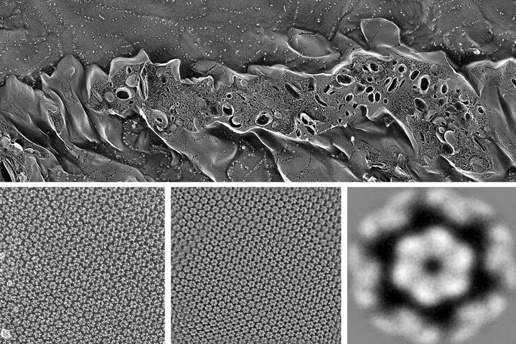
冷冻断裂和冷冻蚀刻是研究柔性膜相关结构(如紧密连接或肠道糖萼)的有用工具。冷冻断裂和冷冻蚀刻是两种互补的方法,通过样品玻璃化来保护目标结构,然后断开冷冻标本以揭示内部结构。冷冻蚀刻是一个后续步骤,在真空下表面冰升华以揭示更多细节。在这些技术中,喷镀金属或碳使样品能够直接在冷冻扫描电子显微镜(SEM)中成像,或作为复制膜在透射电子显微镜(TEM)中成像。这对技术用于研究细胞器、膜、层和乳液,特别适用…
Loading...
探索病毒结构与生命周期
SARS-CoV-2疫情始于2019年12月下旬,随后演变为全球大流行,引发世界范围内抗击COVID-19的斗争。持续发展的电子显微技术提供了大量新应用,使研究人员能够研究病毒结构、感染与复制过程。本网络研讨会将概述这些先进技术,并阐释它们如何揭示病毒感染细胞时引发的复杂变化。我们涵盖了当前用于扫描电镜(SEM)和透射电镜(TEM)的样品制备流程,包括常温、混合及专用冷冻工作流程。
