Webinars

最新市场活动资讯

全部活动

Complete camera overview of EM grid recorded with 3 channels. Inserts displaying the positions, where superresolved 3D confocal images were recorded. 3D renderings of these positions are shown in the zoomed inserts. Fluorescence channels (nuclei by Hoechst, blue; mitochondria by MitoTracker Green, green; lipid Droplets by Bodipy and Crimson Beads, red). Width of a grid square is 90 ?m, width of a grid bar is 35 ?m. Samples kindly provided by Ievgeniia Zagoriy, Mahamid-Group, EMBL Heidelberg, Germany.

从显微镜到电镜:完整的冷冻光电联用工作流程

在题为“多模态玻璃化征程,从实验台到电子显微镜的冷冻关联工作流程”的网络研讨会上,专家团队(Edoardo D'Imprima、Zhengyi Yang、Andreia Pinto 和 Martin…
Developing embryos of different species at different stages during the elongation of their posterior body axis, from left to right in developmental time. The labelled regions in red depict a region of undifferentiated cells called the tailbud, with the corresponding region generated from that tissue shaded in grey. Upper row: lamprey; middle row: catshark; bottom row, zebrafish. This figure has been adapted from the following publication: Steventon, B., Duarte, F., Lagadec, R., Mazan, S., Nicolas, J.-F., & Hirsinger, E. (2016). Species tailoured contribution of volumetric growth and tissue convergence to posterior body elongation in vertebrates. Development, 2016. 143(10):1732-41

如何研究胚胎发育中的基因调控网络

欢迎参加由 Ben Steventon 博士与 Andrea Boni 博士主讲的点播网络研讨会,探索光片显微镜如何革新发育生物学研究。这项先进成像技术能对三维样本进行高速、大体积的活体成像,且光毒性低。通过用户案例了解光片显微镜如何深化我们对肠道类器官与脑类器官发育的认知,并深入解析徕卡显微系统 Viventis Deep 显微镜的技术原理及其在长时间成像中的应用。
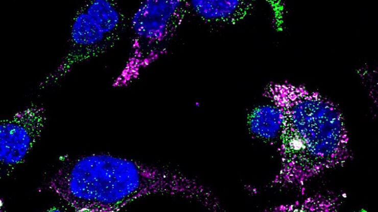
GLP-1 and PYY localized to distinct secretory pools in L-cells.

前沿成像技术用于 GPCR 信号传导

通过这个按需网络研讨会,提升您的药理研究,了解 GPCR 信号传导,并探索旨在理解 GPCR 信号如何转化为细胞和生理反应的尖端成像技术。发现领先的研究,扩展我们对这些关键通路的认识,以寻找新的药物发现途径。
Stripe assay performed on a THUNDER Imager Cell. Courtesy of Maria Carrasquero Ordaz, University of Oxford.

揭示神经元迁移的分子奥秘

研究发育中大脑神经元向生态位迁移可采用多种方法。在本场研讨会中,牛津大学的专家们将展示他们用于阐明神经发育期间神经元向皮层功能层迁移的分子机制的显微技术与实验方法。理解这些过程将有助于更深入地认识健康大脑的发育机制,并可能为神经发育障碍提供更优治疗方案。
Dapi – Nucleus, GFP – Plasma Membrane, Thickness 100µm, 63x objektive, 469 Z planes, 2 channels, THUNDER Imager 3D Cell Culture. Courtesy M.Sc. Dana Krauß, Medical University of Vienna (Austria).

您的 3D 类器官成像和分析工作流程效率如何?

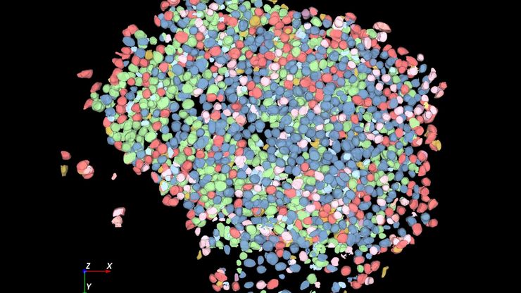
类器官模型已经改变了生命科学研究,但优化图像分析协议仍然是一个关键挑战。本次网络研讨会探讨了类器官研究的简化工作流程,首先是实时的三维细胞培养检查,接下来是高速、高分辨率的三维成像,生成清晰的图像和更纯净的数据,以便对生长速率、细胞迁移和三维细胞相互作用等参数进行准确地人工智能分割和量化,从而实现更深入的洞察。
UC Enuity

通过自动切片改善您的超薄切片工作流程

在不断发展的电镜样品制备领域,保持领先地位至关重要。这个网络研讨会提供了关于超薄切片最新进展的重要见解,这些进展可以显著增强您实验室的能力。
Scroll to top