
History, Developments and Trends of Microscopy in Cancer Research
Cancer is a global disease, with 18 million new cases diagnosed and 10 million cancer-related deaths worldwide in 2020. This burden is set to increase, with a projected increase in cases of ~55% by…
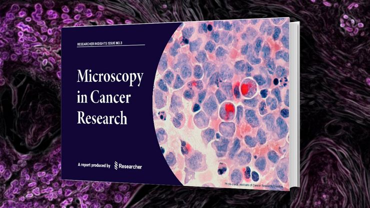
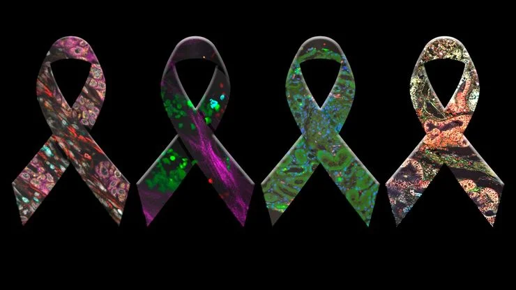
Researchers Insights: Microscopy in Cancer Research
Discover how imaging techniques are driving cancer research forward. In this issue, we present comprehensive multimodal studies using microscopy, as well as new directions in intraoperative cancer…

人工智能与深度视觉蛋白质组学 (DVP) 相结合,推进疾病研究
在这次网络研讨会上,Andreas Mund 博士将介绍深度可视蛋白质组学(DVP)--一种将人工智能驱动的组织空间分辨、非靶向蛋白质组学相结合的尖端平台。他展示了 DVP 如何从最小的、表型匹配的细胞群中识别数千种蛋白质,并在复杂的临床组织样本中生成高分辨率分子图谱,从而在细胞水平上解码疾病机制。
.")
人工智能驱动的乳腺癌研究多重染色成像空间分析工具
乳腺癌(BC)是女性因癌症死亡的主要原因,研究查肿瘤微环境(TME)对于阐明肿瘤进展机制至关重要。利用超多标染色空间蛋白质组学技术系统地绘制肿瘤微环境图谱可以提高精准免疫肿瘤学的能力。在这里,我们将基于人工智能的高倍空间分析应用于BC组织,研究免疫细胞类型和生物标记物,从而深入了解受免疫疗法反应的TME分子机制。
 segmentation used in conjunction with LMD to increase discovery throughput.")
利用激光显微切割发现生物标记物
探索空间蛋白质组学工作流程的潜力,如深度视觉蛋白质组学(DVP),以破译病理机制和发现药物靶点。蛋白质表达、丰度或活性的改变会严重影响细胞功能--通常会导致疾病。值得注意的是,相邻细胞之间的蛋白质组可能存在巨大差异。空间蛋白质组学关注到这种细胞异质性,从而揭示了病理机制。激光显微切割技术(LMD)可获取单细胞进行下游分析,同时保留其空间环境,为空间蛋白质组学奠定了基础。
 and tubulin (magenta), acquired using Viventis Deep. Courtesy of Akanksha Jain, Treutlein Lab ETH-DBSSE Basel (Switzerland).")
如何深入了解类器官和细胞球模型
在本电子书中,您将了解3D细胞培养模型(如类器官和细胞球)成像的关键注意事项。探索创新型显微镜解决方案,来实时记录类器官和细胞球的动态成像过程。

多重成像揭示结肠癌的肿瘤免疫格局
由于抗药性和复发,癌症免疫疗法获益者寥寥无几,而针对癌症免疫周期多个步骤的组合治疗策略可能会改善治疗效果。这项研究表明,高通量空间蛋白质组学可用于识别细胞生物标志物之间的相互作用,并通过绘制肿瘤免疫微环境图来指导精准的组合疗法。
")
如何优化多标成像技术推动3D空间组学发展
本次网络研讨会上,徕卡显微系统的Julia Roberti博士与Luis Alvarez博士将介绍STELLARIS共聚焦平台的全新功能SpectraPlex,该技术可实现超多标三维空间成像。该技术旨在通过实现超多标成像且无需频繁人工干预,从而简化和增强空间生物学应用。

利用空间蛋白质组学工作流程改革研究工作
空间蛋白质组学是《自然-方法》2024 年度方法,正在推动癌症、免疫学等领域的研究进展。通过将定位数据与组织中蛋白质的高通量成像结合起来,研究人员可以发现疾病进展和治疗反应方面的洞察力,从而更好地了解人类生物学。在这里,您可以了解更多有关空间生物学的信息,以及徕卡显微系统的工具如何推动蛋白质生物标记的可视化和分析取得进展。
