

基于激光的视神经再生研究新方法
由于哺乳动物中枢神经系统(CNS)的自我修复能力有限以及传统损伤模型的不一致性,视神经再生是神经生物学的一大挑战。相比之下,爪蟾蝌蚪的视神经在受伤后可以再生,因此是研究轴突再生的分子和细胞机制的理想模型。在本应用说明中,我们展示了如何利用激光显微切割技术(LMD)对蝌蚪的视神经进行精确、一致的横切,从而开发出适合成像、转录组分析和功能恢复研究的高重复性损伤模型。

如何选择合适的测量显微镜
使用测量显微镜,用户可以测量样品特征的二维和三维尺寸,这对检测、质量控制、故障分析和研发&D 至关重要。然而,选择合适的显微镜需要评估应用需求以及显微镜的性能、易用性和灵活性。如今,测量通常以数字方式进行,即使用带有摄像头和软件的显微镜,图像显示在显示器上,而不是通过目镜网线,从而提高了精度和可重复性。使用合适的测量显微镜可靠、快速地分析样品。

罕见疾病 CRISPR 疗法的开发与风险解除
Fyodor Urnov博士和Sadik Kassim博士最初是在ASGCT 2025会议上作这一按需演讲的,演讲的重点是遗传医学中的一个关键挑战:如何将CRISPR疗法从单一疾病解决方案扩展到平台方法,特别是针对罕见的儿科遗传疾病。Urnov 博士展示了由 Matthew Kan 博士领导的创新基因组研究所的工作,这是 IGI-Danaher Beacon for CRISPR Cures…

显微镜测量校准:为什么要校准以及如何校准
显微镜校准可确保用于检测、质量控制 (QC)、故障分析和研发 (R&D) 的测量结果准确一致。本文介绍了校准步骤。使用参照物进行校准可获得可重复的结果,并有助于确保与准则和标准一致。为获得准确一致的结果,建议校准显微镜并定期检查。如有需要,可向校准专家寻求支持。

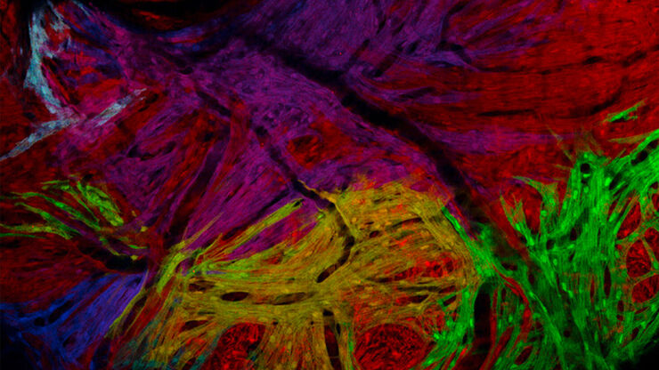
多重成像揭示结肠癌的肿瘤免疫格局
由于抗药性和复发,癌症免疫疗法获益者寥寥无几,而针对癌症免疫周期多个步骤的组合治疗策略可能会改善治疗效果。这项研究表明,高通量空间蛋白质组学可用于识别细胞生物标志物之间的相互作用,并通过绘制肿瘤免疫微环境图来指导精准的组合疗法。
 observed with an Ivesta 3 stereo microscope during fly pushing (sorting of the flies). The scale bar length is 1 mm. Image courtesy of M. Benton, EMBL, Heidelberg, Germany.")
Drosophila(果蝇)研究显微镜使用指南
一个多世纪以来,果蝇(典型的黑腹果蝇)一直被用作模式生物。原因之一是果蝇与人类共享许多与疾病相关的基因。果蝇经常被用于发育生物学、遗传学和神经科学的研究。果蝇的优点包括易于饲养且成本低廉、繁殖速度快、基因组完全测序以及可获得各种基因品系。使用徕卡显微镜可以进行高效的果蝇研究。

神经科学研究指南
神经科学通常需要研究具有挑战性的标本,以更好地了解神经系统和疾病。徕卡显微镜帮助神经科学家深入了解神经元功能。
斑马鱼研究指南
在斑马鱼研究过程中,尤其是在筛选、分类、处理和成像过程中,要想获得最佳结果,看到精细的细节和结构非常重要。他们帮助研究人员为下一步做出正确的决定。徕卡体视显微镜以出色的光学性能和分辨率著称,配备透射光基底和荧光照明,为斑马鱼成像提供了合适的解决方案。高分辨率、色彩保真度和最佳对比度使研究人员能够做出具有洞察力的决策。

利用快速高对比度成像改进斑马鱼-胚胎筛查
通过这篇文章,您可以了解如何利用 DM6 B 显微镜的高速、高对比度成像技术促进转基因斑马鱼胚胎的筛选,从而确保发育生物学研究的准确定位。
