STELLARIS DLS
共聚焦显微镜
产品
首页
Leica Microsystems
STELLARIS DLS 数字光片显微镜
重新定义光片显微镜
阅读我们的最新文章

基于人工智能的表型药物筛查解决方案
本次网络研讨会将全面介绍使用三维细胞培养进行表型药物筛选所遇到的问题、可能的解决方案及规划与执行策略。

STELLARIS 共聚焦显微镜平台的虚拟现实展示
In this webinar, you will discover how to perform 10-color acquisition using a confocal microscope. The challenges of imaged-based approaches to identify skin immune cells. A new pipeline to assess…
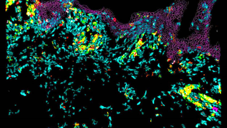
免疫细胞在组织样品中的共聚焦成像
在本次网络研讨会中,您将探索如何使用共聚焦显微镜对组织样品进行10色成像,并了解这一技术如何有助于评估皮肤免疫状况。

利用DLS对细胞球中的抗癌药物摄取进行成像
细胞球3D细胞培养模型模拟了活组织的生理和功能,使其成为研究肿瘤形态和筛选抗癌药物的有用工具。药物AZD2014是一种公认的哺乳动物雷帕霉素靶蛋白(mTOR)通路抑制剂[1]。mTOR的异常激活会促进肿瘤生长和转移,导致AZD2014进入临床试验作为抗癌分子。其具体的抗肿瘤机制尚不清楚。

模式生物研究
模式生物是研究人员用来研究特定生物学过程的物种。 它们具有与人类相似的遗传特征,通常用于遗传学、发育生物学和神经科学等研究领域。 选择模式生物的原因通常是它们在实验室环境中易于保持和繁殖、生成周期短,或能够产生突变体来研究某些性状或疾病。

利用光片显微镜改进三维细胞生物学工作流程
了解癌症发生过程中的亚细胞机制对于癌症治疗至关重要。常见的细胞模型涉及作为单层生长的癌细胞。然而,这种方法忽视了肿瘤细胞与其周围微环境之间的三维相互作用。为了贴近自然环境理解恶性肿瘤的发展和进程,对癌症微环境的详细表征至关重要。
![3D glomeruli in a portion of an ECi-cleared kidney scanned by light sheet microscopy. Courtesy of Prof. Norbert Gretz, Medical Faculty Mannheim, University of Heidelberg [1].](/fileadmin/_processed_/d/d/csm_DLS-Sample-Preparation-Intr_915e0fd7c2.jpg "3D glomeruli in a portion of an ECi-cleared kidney scanned by light sheet microscopy. Courtesy of Prof. Norbert Gretz, Medical Faculty Mannheim, University of Heidelberg [1].")
使用安装框架进行光片样品准备
样品处理通常是光片显微镜研究中的一个关键话题。徕卡显微系统的TCS SP8 DLS将光片技术集成到倒置共聚焦平台中,因此可以利用关于样品安装和XY-stage功能的一般原则。本文将描述一组安装框架,这些框架不仅允许准备更多的样品,尤其是在使用诸如BABB(苯甲醇苯甲酸酯)等潜在有害的安装介质时,亦具有广泛的适用性。

使用旋转设备进行光片样本安装
TCS SP8 DLS 显微镜采用了一种创新的设计理念,将光片显微技术集成到共聚焦显微镜中。得益于其独特的光学架构,样本可以像进行标准共聚焦成像一样,安装在标准玻璃底培养皿上。与传统的样本安装程序相比,这一过程只需进行少量调整。

使用 U 形玻璃毛细管进行样品装载
徕卡显微系统的DLS显微镜系统是一种创新概念,将光片显微技术集成到共聚焦平台中。由于其独特的光学结构,样本可以安装在标准玻璃底培养皿上,与传统的安装程序相比,几乎不需要或只需很少的适应。在这里,我们介绍了一种便捷的方法,能够快速准备样本以进行光片成像。

共聚焦成像和光片成像
光学成像仪器可以放大微小物体,聚焦遥远星体,揭示肉眼看不见的细节。但是,它有一个众所周知且令人烦恼的问题:景深有限。我们的眼睛(也是一种光学成像装置)也有同样的困扰,但我们的大脑在信号到达意识认知之前会巧妙地移除所有不在焦点上的信息。
