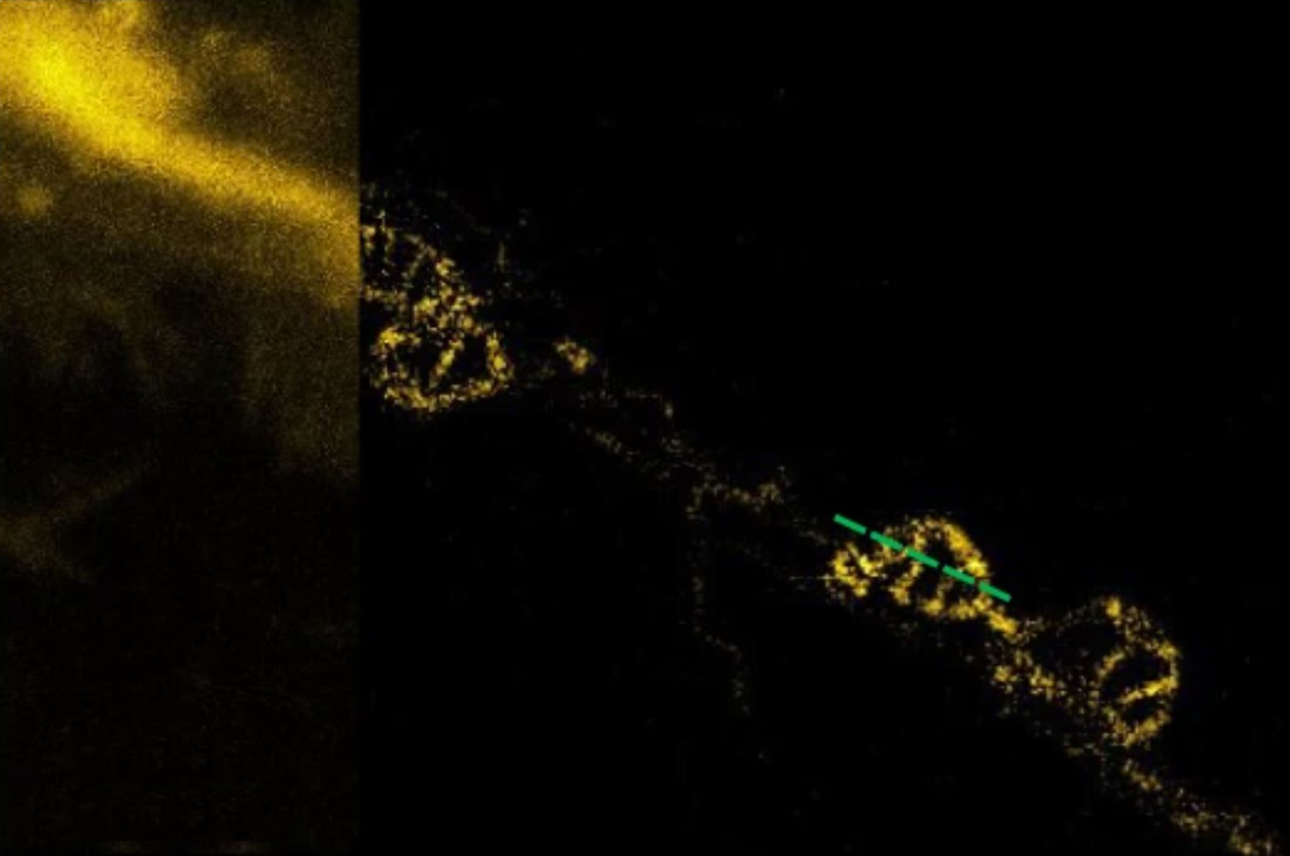
![[Translate to chinese:] STED imaging of mitochondria revealed cristae dynamics during fusion. A comparison of confocal (left) and STED (right) imaging. Mitochondrial_dynamics_quantitatively_revealed_by_STED_nanoscopy_teaser.JPG](/fileadmin/_processed_/d/8/csm_Mitochondrial_dynamics_quantitatively_revealed_by_STED_nanoscopy_teaser_3a80e0f008.jpg "[Translate to chinese:] STED imaging of mitochondria revealed cristae dynamics during fusion. A comparison of confocal (left) and STED (right) imaging.")
Yang X., Yang Z., Wu Z., He Y., ShanC., Chai P., Ma C., Tian M., Teng J., Jin D., Yan W., Das P., Qu J. & Xi P.:
Mitochondrial dynamics quantitatively revealed by STED nanoscopy with an enhanced squaraine variant probe
Nature Communications volume 11, Article number: 3699 (2020)

相关文章
-
![[Translate to chinese:] Multicolor fixed STED image. Inner ear section, mouse, TauSTED Xtend 589 on AF488 and TauSTED Xtend 775 on AF633-Phalloidin. Sample courtesy of Dennis Derstrof, Klinik für Hals-, Nasen und Ohrenheilkunde, Universität Marburg & Prof. Dr. Dominik Oliver aus dem Institut für Physiologie und Pathophysiologie, Abteilung für Neurophysiologie, Universität Marburg.](/fileadmin/_processed_/c/a/csm_Inner_ear_section_mouse_multicolor_fixed_TauSTED_Xtend_3410e9781c.jpg "[Translate to chinese:] Multicolor fixed STED image. Inner ear section, mouse, TauSTED Xtend 589 on AF488 and TauSTED Xtend 775 on AF633-Phalloidin.")
细胞活成像的纳米级扩展
新的STED显微技术方法——TauSTED Xtend,使得在纳米级别下对活体完整样本进行扩展多色成像成为可能。通过结合空间和寿命信息,TauSTED…
Mar 05, 2024Read article -
![[Translate to chinese:] Multicolor TauSTED Xtend 775 for Cell Biology applications that require nanoscopy resolution for multiple cellular components. Cells showing vimentin fibrils (AF 594), actin network (ATTO 647N), and nuclear pore basket (CF 680R). Sample courtesy of Brigitte Bergner, Mariano Gonzales Pisfil, Steffen Dietzel, Core Facility Bioimaging, Biomedical Center, Ludwig-Maximilians-University, Munich, Germany.](/fileadmin/_processed_/5/4/csm_Triple_color_fixed_sample_TauSTED_Xtend_2_3_c7d82e8a27.jpg "[Translate to chinese:] Multicolor TauSTED Xtend 775 for Cell Biology applications that require nanoscopy resolution for multiple cellular components. Cells showing vimentin fibrils (AF 594), actin network (ATTO 647N), and nuclear pore basket (CF 680R).")
STED样品制备指南
这份指南旨在帮助用户优化受激发射损耗(STED)纳米成像的样品制备,特别是在使用徕卡微系统的STED显微镜时。它提供了单色STED成像用荧光标记的概述,并对其性能进行了评级。
Mar 05, 2024Read article -
![[Translate to chinese:] Five-color FLIM-STED](/fileadmin/_processed_/a/3/csm_5-color_FLIM-STED_0cd701139b.jpg "[Translate to chinese:] Five-color FLIM-STED")