
.")
利用光片显微技术聚焦三维长时程成像
长时程三维成像揭示了复杂的多细胞系统是如何生长和发育的,以及细胞是如何随着时间的推移而移动和相互作用的,从而揭示了发育、疾病和再生方面的重要知识。光片显微镜一次只照射样品的一个薄片,大大减少了光损伤,保护了样品的活性。这种温和的高速技术可在数小时甚至数天内提供清晰的体数据,使研究人员能够实时捕捉生物学的发展过程。

Capturing Developmental Dynamics in 3D
This application note showcases how the Viventis Deep dual-view light sheet microscope was successfully used by researchers for exploring high-resolution, long-term imaging of 3D multicellular models…
。红色标记区域显示未分化细胞区(尾芽),灰色阴影表示该组织生成的对应区域。上图:七鳃鳗;中图:猫鲨;下")
如何研究胚胎发育中的基因调控网络
欢迎参加由 Ben Steventon 博士与 Andrea Boni 博士主讲的点播网络研讨会,探索光片显微镜如何革新发育生物学研究。这项先进成像技术能对三维样本进行高速、大体积的活体成像,且光毒性低。通过用户案例了解光片显微镜如何深化我们对肠道类器官与脑类器官发育的认知,并深入解析徕卡显微系统 Viventis Deep 显微镜的技术原理及其在长时间成像中的应用。
.")
双视野光片显微镜,适用于大型多细胞系统
展示复杂多细胞系统的动态是生物学中的一个基本目标。为了应对在大型时空尺度上进行活体成像的挑战,作者在《自然·方法》杂志上发表的一篇论文中介绍了一种开放式多样本双视野光片显微镜。研究发现,Viventis LS2 Live显微镜在以单细胞分辨率成像大型样本方面取得了显著进展。

基于人工智能的表型药物筛查解决方案
本次网络研讨会将全面介绍使用三维细胞培养进行表型药物筛选所遇到的问题、可能的解决方案及规划与执行策略。
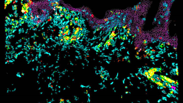
免疫细胞在组织样品中的共聚焦成像
在本次网络研讨会中,您将探索如何使用共聚焦显微镜对组织样品进行10色成像,并了解这一技术如何有助于评估皮肤免疫状况。
 and mito OM (red) in a live U2OS cell")
多色四维超分辨光片显微镜
人工智能显微术研讨会主要关注和讨论显微术和生物医学成像领域的最新人工智能技术和工具。在该科学演示中,Yuxuan Zhao展示了如何通过渐进式深度学习策略并结合“双环调制的SPIM”设计改善活细胞中的细胞器三维成像。

利用DLS对细胞球中的抗癌药物摄取进行成像
细胞球3D细胞培养模型模拟了活组织的生理和功能,使其成为研究肿瘤形态和筛选抗癌药物的有用工具。药物AZD2014是一种公认的哺乳动物雷帕霉素靶蛋白(mTOR)通路抑制剂[1]。mTOR的异常激活会促进肿瘤生长和转移,导致AZD2014进入临床试验作为抗癌分子。其具体的抗肿瘤机制尚不清楚。
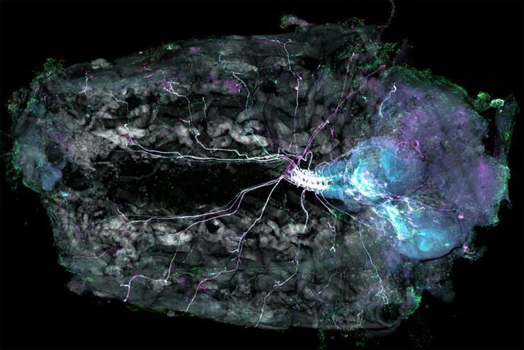
Understanding Motor Sequence Generation Across Spatiotemporal Scales
We have developed a microscopy-based pipeline to characterize a developmentally critical behavior at the pupal stage of development, called the ecdysis sequence. We study brain-wide neuronal activity…
