

神经科学显微镜面临哪些挑战?
显微镜是神经科学研究领域的强大工具。不过,当涉及到对神经过程进行成像以及使用不同的样品类型(例如厚神经组织或脑类器官)时,科研人员可能会面临到很多挑战。这本30页的电子书包含众多真实的案例,以讨论我们最常见到的一些挑战,同时展示了如何使用THUNDER 成像技术克服这些挑战。

铁代谢在癌症进展中的作用
铁代谢在癌症发展和演进过程中发挥着重要作用,可以调节免疫反应了解铁离子如何影响癌症和免疫系统,有助于开发新的癌症治疗方法。

超越反卷积
宽场荧光显微镜通常用于视觉呈现生命科学样本中的结构并获取重要信息。利用荧光蛋白或染料,以高度特异性的方式标记离散的样本部分。为了充分了解某种结构,可能需要以三维方式呈现,但这会对使用显微镜带来某些挑战。
 stained to show the nucleus")
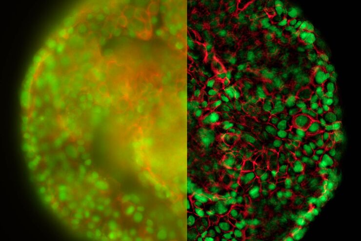
使用Mica的人工智能显微镜软件进行 3D 空间分析
本期MicaCam为您提供切实的建议,教您从显微镜图像中提取可发表级别的分析结果。本期的特邀嘉宾来自徕卡显微系统的Luciano Lucas,他将为大家展示如何使用MICA的AI赋能软件进行图像分析。他将深度分析两张MICA的3D成像,探究不同可见生物元素之间的空间关系。本期的最后将会介绍如何创作高保真视频动画以及其他可用于发表文章的结果。

High-resolution 3D Imaging to Investigate Tissue Ageing
Award-winning researcher Dr. Anjali Kusumbe demonstrates age-related changes in vascular microenvironments through single-cell resolution 3D imaging of young and aged organs.

利用光片显微镜改进三维细胞生物学工作流程
了解癌症发生过程中的亚细胞机制对于癌症治疗至关重要。常见的细胞模型涉及作为单层生长的癌细胞。然而,这种方法忽视了肿瘤细胞与其周围微环境之间的三维相互作用。为了贴近自然环境理解恶性肿瘤的发展和进程,对癌症微环境的详细表征至关重要。
清晰对比、无雾的 3D 样本实时图像
历史上,宽场显微镜并不适合对大样本/标本体积进行成像。图像背景(BG)主要来源于观察样本的失焦区域,显著降低了成像系统的对比度、有效动态范围和最大可能的信噪比(SNR)。记录的图像显示出典型的雾霭,并且在许多情况下,无法提供进一步分析所需的细节水平。处理厚三维样本的研究人员要么使用替代显微镜方法,要么尝试通过后处理一系列图像来减少雾霭。

带有全自动连续切片功能的高分辨率序列断层成像
本报告描述了利用全自动连续切片方案通过序列断层成像对高分辨率三维亚细胞结构分析进行优化,在基底上实现高切片密度。
高分辨率共聚焦显微镜的 BABB 清洗和成像
Multipohoton microscopy experiment using Leica TCS SP8 MP and Leica 20x/0.95 NA BABB immersion objective.
Understanding kidney microanatomy is key to detecting and identifying early events in kidney…
