揭示神经元迁移的分子奥秘
研究发育中大脑神经元向生态位迁移可采用多种方法。在本场研讨会中,牛津大学的专家们将展示他们用于阐明神经发育期间神经元向皮层功能层迁移的分子机制的显微技术与实验方法。理解这些过程将有助于更深入地认识健康大脑的发育机制,并可能为神经发育障碍提供更优治疗方案。
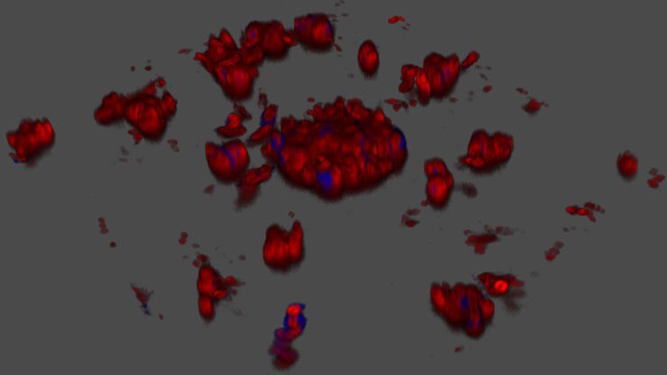
探索微生物世界:三维食品基质中的空间相互作用
Micalis 研究所是与 INRAE、AgroParisTech 和巴黎萨克雷大学合作的联合研究单位。其使命是开发食品微生物学领域的创新研究,以促进健康。在这一系列视频中,Micalis…
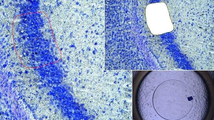
激光微切割(LMD)促进的分子生物学分析
使用激光微切割(LMD)提取生物分子、蛋白质、核酸、脂质和染色体,以及提取和操作细胞和组织,可以深入了解基因和蛋白质的功能。它在神经生物学、免疫学、发育生物学、细胞生物学和法医学等多个领域有广泛应用,例如癌症和疾病研究、基因改造、分子病理学和生物学。LMD 也有助于研究蛋白质功能、分子机制及其在转导途径中的相互作用。
使用空间多重化探测人类阿尔茨海默病皮层切片
阿尔茨海默病(AD)是最常见的神经退行性疾病,其特征是认知功能的逐渐下降。对 AD 大脑的空间分析可能揭示细胞关系,从而促进对疾病病因的更好理解。本研究捕捉了 AD 皮层组织成分的全球概述,并强调了 Cell DIVE 成像的简化工作流程,从数据采集到使用 Aivia 软件的基于人工智能的分析,最终实现更快的洞察。
空间代谢组学:探索肿瘤复杂性和治疗见解
在癌症研究中,理解肿瘤细胞与其微环境之间的相互作用至关重要,因为肿瘤微环境显著影响肿瘤进展。空间代谢组学是一种由研究人员开发的新方法,用于研究这一复杂性。通过揭示肿瘤微环境中的空间变化,该方法提供了对其多样化成分及其组织的宝贵见解。这些见解不仅影响临床结果,还为治疗反应提供信息,为个性化治疗策略铺平道路。
利用子宫内膜类器官推进子宫再生疗法
康教授团队致力于研究决定子宫微环境的关键因素,该环境对胚胎着床和妊娠维持至关重要。他们正为罹患阿什曼综合征等子宫内膜疾病的患者开发恢复子宫内膜功能的新型治疗策略。通过将 3D 子宫内膜类器官移植至小鼠模型,该团队揭示了子宫内膜强大的再生能力的细胞与分子机制。本次访谈将深入探讨其团队的研究内容及Mica在研究中所发挥的重要作用。
基于激光显微切割的稀疏细胞脂质组学分析
通过高覆盖率靶向脂质组学分析稀疏细胞,深入探讨细胞复杂性。这种先进的方法结合了激光显微切割(LMD)和液相色谱-质谱/质谱(LC-MS/MS),揭示了单细胞水平的代谢变化,阐明了糖尿病和肥胖等疾病。通过采用激光显微切割(LMD)获得无污染样本,并使用 SCIEX 7500 系统提高灵敏度,该方法成功检测到 285…
您的 3D 类器官成像和分析工作流程效率如何?
类器官模型已经改变了生命科学研究,但优化图像分析协议仍然是一个关键挑战。本次网络研讨会探讨了类器官研究的简化工作流程,首先是实时的三维细胞培养检查,接下来是高速、高分辨率的三维成像,生成清晰的图像和更纯净的数据,以便对生长速率、细胞迁移和三维细胞相互作用等参数进行准确地人工智能分割和量化,从而实现更深入的洞察。
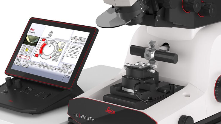
通过自动切片改善您的超薄切片工作流程
在不断发展的电镜样品制备领域,保持领先地位至关重要。这个网络研讨会提供了关于超薄切片最新进展的重要见解,这些进展可以显著增强您实验室的能力。


