Viventis
光片显微镜
产品
首页
Leica Microsystems
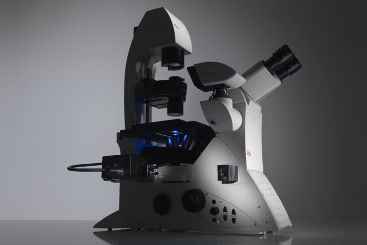
Viventis 双视野光片荧光显微镜
以完整的深度揭开生命的奥秘
阅读我们的最新文章
Guide to Live-Cell Imaging
For a wide range of applications in various research fields of life science, live-cell imaging is an indispensable tool for visualizing cells in a state as close to in vivo, i.e. living and active, as…
Factors to Consider When Selecting a Research Microscope
An optical microscope is often one of the central devices in a life-science research lab. It can be used for various applications which shed light on many scientific questions. Thereby the…
利用光片显微技术聚焦三维长时程成像
长时程三维成像揭示了复杂的多细胞系统是如何生长和发育的,以及细胞是如何随着时间的推移而移动和相互作用的,从而揭示了发育、疾病和再生方面的重要知识。光片显微镜一次只照射样品的一个薄片,大大减少了光损伤,保护了样品的活性。这种温和的高速技术可在数小时甚至数天内提供清晰的体数据,使研究人员能够实时捕捉生物学的发展过程。
如何深入了解类器官和细胞球模型
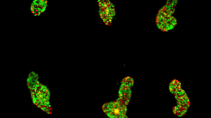
在本电子书中,您将了解3D细胞培养模型(如类器官和细胞球)成像的关键注意事项。探索创新型显微镜解决方案,来实时记录类器官和细胞球的动态成像过程。
来捕捉发育动态的3D成像
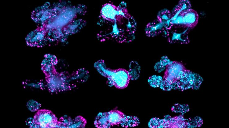
本应用说明展示了研究人员如何成功利用 Viventis Deep 双视角光片显微镜探索3D多细胞模型(包括有机体、球形体和胚胎)的高分辨率长期成像,从而为发育生物学和疾病研究带来新的可能性。
Drosophila(果蝇)研究显微镜使用指南
一个多世纪以来,果蝇(典型的黑腹果蝇)一直被用作模式生物。原因之一是果蝇与人类共享许多与疾病相关的基因。果蝇经常被用于发育生物学、遗传学和神经科学的研究。果蝇的优点包括易于饲养且成本低廉、繁殖速度快、基因组完全测序以及可获得各种基因品系。使用徕卡显微镜可以进行高效的果蝇研究。
神经科学研究指南
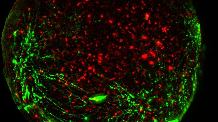
神经科学通常需要研究具有挑战性的样本,以便更好地了解神经系统和疾病。徕卡显微镜可帮助神经科学家深入了解神经元的功能。
如何研究胚胎发育中的基因调控网络
欢迎参加由 Ben Steventon 博士与 Andrea Boni 博士主讲的点播网络研讨会,探索光片显微镜如何革新发育生物学研究。这项先进成像技术能对三维样本进行高速、大体积的活体成像,且光毒性低。通过用户案例了解光片显微镜如何深化我们对肠道类器官与脑类器官发育的认知,并深入解析徕卡显微系统 Viventis Deep 显微镜的技术原理及其在长时间成像中的应用。
双视野光片显微镜,适用于大型多细胞系统
展示复杂多细胞系统的动态是生物学中的一个基本目标。为了应对在大型时空尺度上进行活体成像的挑战,作者在《自然·方法》杂志上发表的一篇论文中介绍了一种开放式多样本双视野光片显微镜。研究发现,Viventis LS2 Live显微镜在以单细胞分辨率成像大型样本方面取得了显著进展。
下载活细胞成像指南
在生命科学研究中,活细胞成像是一种不可或缺的工具,可用于观察细胞的活体状态。这本电子书回顾了为确保成功进行活细胞成像而需要考虑的各种重要因素。
模式生物研究
模式生物是研究人员用来研究特定生物学过程的物种。 它们具有与人类相似的遗传特征,通常用于遗传学、发育生物学和神经科学等研究领域。 选择模式生物的原因通常是它们在实验室环境中易于保持和繁殖、生成周期短,或能够产生突变体来研究某些性状或疾病。
活细胞成像简介
了解复杂且快速变化的细胞动力学是深入探索生物进程的重要一步。因此,现代生命科学研究越来越需要关注于在分子水平上实时发生的生理事件。
Biopharma
For biopharma, Leica solutions help accelerate drug discovery, enhance cellular analysis, and support data integrity that meets regulations.
应用领域
类器官和3D细胞培养
生命科学研究中最令人振奋的最新进展之一是3D细胞培养系统的发展,例如类器官、球状体或器官芯片模型。 3D细胞培养物是一种人工环境,在这种环境中,细胞能够在三维空间中生长并与周围环境相互作用。 这些环境条件与它们在体内的情况相似。

