倒置光学显微镜

徕卡显微系统公司的倒置显微镜旨在满足生命科学、材料科学和工业应用的严格要求。

用于生命科学的倒置显微镜

先进的生命科学研究需要适用于活细胞或组织培养等各种应用的先进成像解决方案。 徕卡倒置显微镜提供高性能,帮助您推进研究。 倒置显微镜提供易于使用的软件、高分辨率的数码相机和优秀的 LED 照明,能够灵活满足您的实验室需求。

用于材料科学和工业制造的倒置显微镜

使用徕卡模块化倒置显微镜快速高效地进行材料检验和分析。 高性能光学系统、直观的控制界面和易于使用的软件,可最大限度地减少全面培训的需要,提高您的工作效率。

是否需要帮助?

请联系我们,获取满足您的需求和预算的合适的倒置显微镜的专业建议。

您的应用是什么?

根据您的应用需求确定最适合的倒置显微镜。 一些倒置复合显微镜和倒置荧光显微镜比其他显微镜更能满足特定应用的需求。 徕卡倒置显微镜采用模块化设计,可为特定应用定制解决方案。

需要查看哪些类型的样本?

无论是观察生命科学研究中的样本,例如细胞或组织,还是材料生产和分析,徕卡倒置显微镜解决方案都能提供所需的光学分辨率、对比度和图像质量。 此外,利用倒置显微镜配件(如物镜、照明系统和数码相机)以及 Leica Application Suite 软件,可以进一步定制和优化解决方案,以满足特定应用需求。

倒置显微镜的预算是多少?

定制倒置显微镜的成本可能更高,但可以提高生产效率。 由于部件和配件种类繁多,倒置显微镜几乎可以针对任何应用进行优化。 

倒置显微镜与正置显微镜的区别是什么? 倒置显微镜可以提供那些优势?

对于倒置显微镜,物镜位于载物台下方,而样本位于物镜上方。 正置显微镜的情况正好相反。 使用倒置显微镜:

  1. 与正置相比,自由度更高。 由于样本位于物镜上方,因此,工作距离更大,可对大型和重型样本进行成像。 
  2. 倒置显微镜更容易观察细胞和组织培养物,因为细胞下沉或粘附在透明培养皿或烧瓶的底部(注满水溶液)。 由于需要水浸物镜,因此很难从上方使用直立显微镜看到很多东西。 
  3. 可以在更短时间内查看更多样本。 只需将样本放在载物台上,聚焦表面一次,然后对其成像。 所有其他样本的焦距保持不变。 
  4. 物镜撞入样本的风险要小得多。 由于物镜位于载物台下方,因此,这个事实有助于极大降低物镜撞入样本的风险。 
  5. 节省样本制备的时间和成本。 只需制备样本的一侧,即可更轻松地对大型样本成像。 


有关详情,请参阅科学实验室文章: 
工业应用中倒置显微镜相比正置显微镜的五个优点

related articles

阅读我们的最新文章

作为徕卡显微系统有限公司 的信息门户,ScienceLab (徕卡课堂) 提供许多关于显微技术主题的科研和教学材料。其内容宗旨在于为初学者、有经验的从业者和科学家等的日常工作和实验提供支持。

More Articles

Final Segmentation of organelles in Trichomonas species. Magenta – costa, light blue – hydrogenosomes, turquoise – ER, red – vacuoles, yellow – axostyle, green – Golgi apparatus.  Sample courtesy of Isabelle Guerin-Bonne, Low Kay En, Electron Microscopy Unit, Yong Loo Lin School of Medicine, National University of Singapore. Scale bar: 1 µm.

Volume EM and AI Image Analysis

The article outlines a detailed workflow for studying biological tissues in three dimensions using volume-scanning electron microscopy (volume-SEM) combined with AI-assisted image analysis. The focus…
Image of roundworm C. elegans acquired with a M205 FA fluorescence automated stereo microscope in combination with Rottermann contrast. Areas labelled with mCherry are seen as reddish purple.

A Guide to C. elegans Research – Working with Nematodes

Efficient microscopy techniques for C. elegans research are outlined in this guide. As a widely used model organism with about 70% gene homology to humans, the nematode Caenorhabditis elegans (also…
Dr. Nordmann in conversation with Dr. Falk Schlaudraff, Manager Product Management Uprights (widefield/compound) at Leica Microsystems

How a Breakthrough in Spatial Proteomics Saved Lives

Toxic epidermal necrolysis (TEN) is a rare but devastating reaction to common medications like antibiotics or gout treatments. It begins innocuously, often as a rash, but can escalate rapidly into…
Dr. Andrew Huang, Baylor College of Medicine, in the operating room (OR) performing ear, nose and throat (ENT) surgery using the MyVeo surgical visualization headset.

A Microvascular Surgeon’s View: How MyVeo Transforms Visualization

In this article, Dr. Andrew T. Huang, MD, FACS, otolaryngologist and a head and neck reconstructive surgeon, shares how digital 3D surgical visualization with the MyVeo headset from Leica Microsystems…

A Novel Laser-Based Method for Studying Optic Nerve Regeneration

Optic nerve regeneration is a major challenge in neurobiology due to the limited self-repair capacity of the mammalian central nervous system (CNS) and the inconsistency of traditional injury models.…
Fluorescence microscopy of sectioned tissue, showing the interface between the extensor digitorum longus muscle and the common peroneal nerve in the adult rat. Regenerative peripheral nerve interface (RPNI) at 2 weeks. Image acquired using Mica. Stained for nuclei (blue), neurofilaments (green) and S100B (red). Image courtesy of Dr. Aaron Lee, Department of Bioengineering (Lab of Dr. Rylie Green), Imperial College London.

How to Image Axon Regeneration in Deep Muscle Tissue

This study highlights Dr. Aaron Lee’s research on mapping nerve regeneration in muscle grafts post-amputation. Limb loss often leads to reduced quality of life, not only from tissue loss but also due…
5 hour time-lapse maximum intensity projection of a zebrafish embryo along the z-axis at 3 days post fertilization. Left: microglia cells. Right: bright field channel. Courtesy of Prof. Francesca Peri, University of Zurich, Switzerland.

Capturing Developmental Dynamics in 3D

This application note showcases how the Viventis Deep dual-view light sheet microscope was successfully used by researchers for exploring high-resolution, long-term imaging of 3D multicellular models…
Some 2D measurements, e.g., lengths and areas, made on a PCB sample with a Leica measurement microscope using the Enersight software.

How to Select the Right Measurement Microscope

With a measurement microscope, users can measure the size and dimensions of sample features in both 2D and 3D, something crucial for inspection, QC, failure analysis, and R&D. However, choosing the…
这些图像说明,要捕捉特定细胞中的所有 gH2Ax 病灶并进行精确计数,用多个三维光切片方法实现。

罕见疾病 CRISPR 疗法的开发与风险解除

Fyodor Urnov博士和Sadik Kassim博士最初是在ASGCT 2025会议上作这一按需演讲的,演讲的重点是遗传医学中的一个关键挑战:如何将CRISPR疗法从单一疾病解决方案扩展到平台方法,特别是针对罕见的儿科遗传疾病。Urnov 博士展示了由 Matthew Kan 博士领导的创新基因组研究所的工作,这是 IGI-Danaher Beacon for CRISPR Cures…
Example of calibrating a microscope at a higher magnification value using a stage micrometer.

显微镜测量校准:为什么要校准以及如何校准

显微镜校准可确保用于检测、质量控制 (QC)、故障分析和研发 (R&D) 的测量结果准确一致。本文介绍了校准步骤。使用参照物进行校准可获得可重复的结果,并有助于确保与准则和标准一致。为获得准确一致的结果,建议校准显微镜并定期检查。如有需要,可向校准专家寻求支持。

材料科学与检测

出色的样品制备和成像方法是可靠、准确地检测材料和观察细节的关键。与正置显微镜相比,倒置显微镜能让您以更高效的方式实现这一目标。倒置显微镜可为您提供高质量的光学镜组和智能自动化,从而实现理想的工作流程。

了解详情

生命科学研究

徕卡(Leica)生命科学显微镜凭借先进的创新和专业技术能力,支持观察、测量和分析微结构的成像要求。徕卡显微系统对科学应用领域的高度关注,使徕卡显微系统的用户始终保持领先位置

了解详情

倒置显微镜常见问题解答

Show answer 倒置显微镜的工作原理是什么?

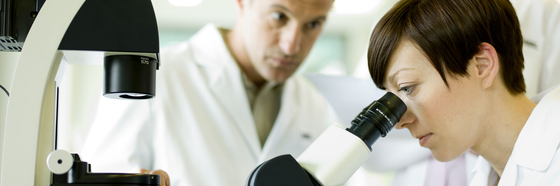
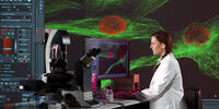
使用倒置显微镜,可以从下方观察样本,因为它们的光学器件放置在样本台下方。 这与从上方观察样本的正置显微镜相反。 倒置显微镜通常用于在生命科学研究中观察细胞和组织培养物,因为细胞沉降或粘附在装满水溶液(生长培养基)的透明培养皿或烧瓶的底部,而从上面无法看到很多细胞和培养物。 倒置显微镜在材料应用中也越来越受欢迎。 有关详情,请参阅科学实验室文章:

光学显微镜——一些基础知识
工业应用中倒置显微镜相比正置显微镜的五个优点

Show answer 倒置显微镜配有哪些类型的配件? 需要单独购买什么?

徕卡倒置显微镜采用模块化设计,以最适合指定需求或应用的配置交付。 如果以后需要改变,可以通过添加可用配件来升级工作站。

Show answer 是否有多个用户使用一台显微镜? 哪些徕卡倒置光学显微镜可以设置个人用户的偏好?

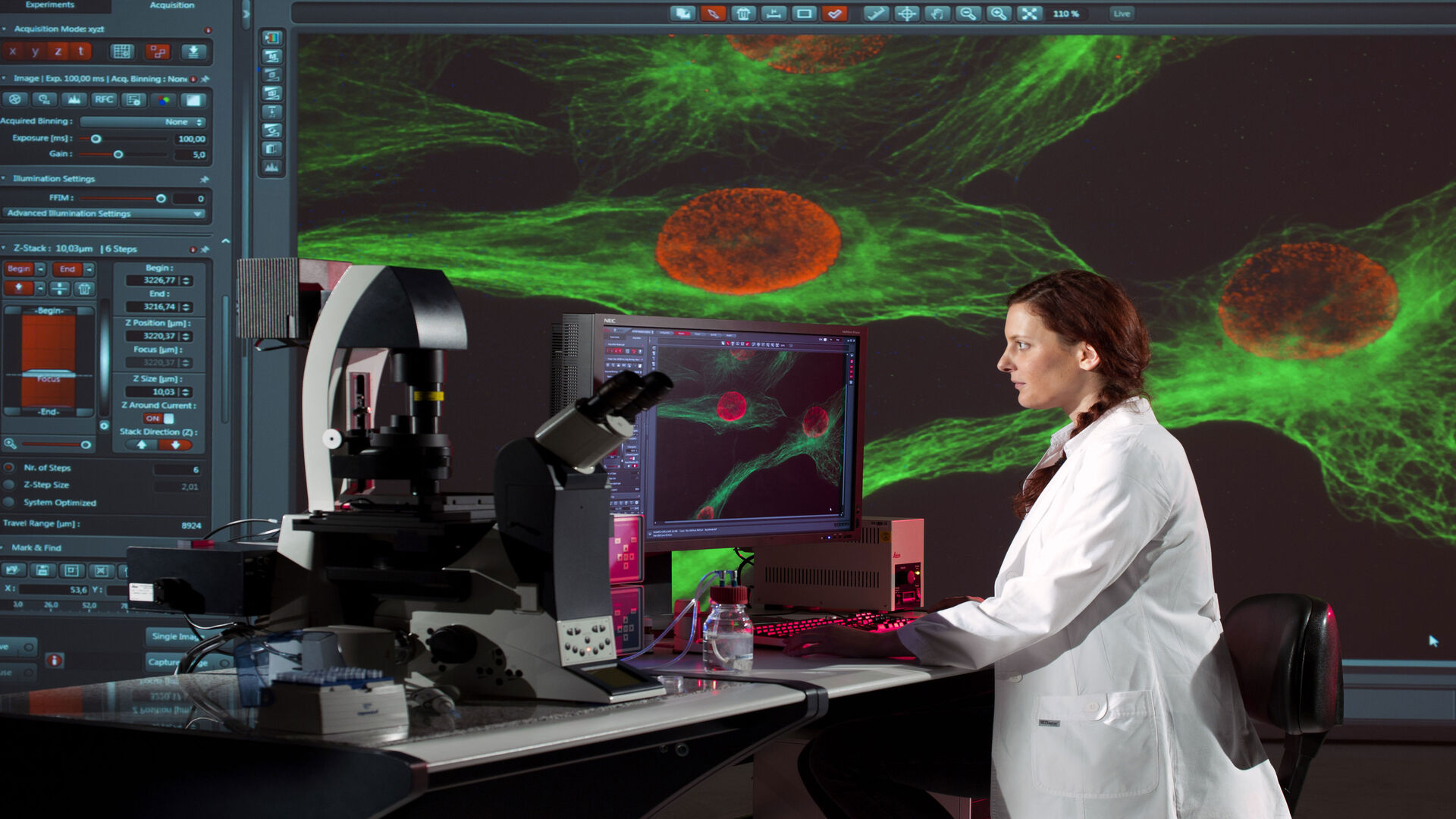
徕卡显微系统公司提供免费的 Store & Recall 软件(适用于工业 LAS X 软件平台),可以根据每个用户的要求和需求来定制显微镜功能。 使用该软件,还能够恢复随采集的图像一起保存的所有系统设置。

Show answer 经过校准且可比较的图像是必不可少的。 推荐使用哪些类型的倒置复合显微镜来满足这一需求?

我们所有的编码显微镜解决方案都提供校准和图像对比。 此外,徕卡显微系统公司提供免费的 Store & Recall 软件(适用于工业 LAS X 软件平台),支持恢复随采集的图像一起保存的所有系统设置。

Show answer 是否需要一台计算机来操作徕卡倒置显微镜?

不用。 通过安装 FLEXACAM C1 摄像头,可以直接将图像保存在 IT 网络服务器或 USB 媒体上。 还可以通过网络使用电子邮件发送图像,无需使用 PC。

Show answer 我需要快速记录并与团队成员分享结果。 推荐的最佳解决方案是什么?

使用 FLEXACAM C1 摄像头,可以直接将图像保存在 IT 网络服务器或 USB 媒体上。 也可以通过电子邮件发送图像,而无需使用计算机。

Show answer 有哪些模块化配件可以扩展我的显微镜的使用范围?

有很多配件。 请联系当地的徕卡销售代表。

Show answer 符合人体工程学的配件如何减少长时间工作造成的重复性劳损?

徕卡显微镜有很多符合人体工程学的配件。 请联系当地的徕卡销售代表,了解详细信息或访问: 用于体视显微镜的人体工程学配件

Show answer Leica Application Suite (LAS) X 软件是否兼容 Windows 和 MAC? Linux 呢?

LAS X 软件只能在 Windows 系统上运行,但对于 MAC 系统,我们提供了一个名为 Leica Acquire 的专用软件,可以从 Apple 商店免费下载:https://apps.apple.com/it/app/leica-acquire/id733706983?mt=12

不过,没有用于 Linux 的软件。

Show answer 徕卡显微镜是否只与 Leica Application Suite 软件兼容? 是否可以与第三方软件集成?

是的,可以使用第三方软件:https://www.splashtop.com/classroom

Show answer 除了徕卡相机之外,我可以在徕卡显微镜上安装第三方 SLR/DSLRc 相机吗?

我们为所有兼容 C-mount 的相机提供适配器。

Scroll to top