STELLARIS STED
共聚焦显微镜
产品
首页
Leica Microsystems
STELLARIS STED
更快了解真实的世界
阅读我们的最新文章
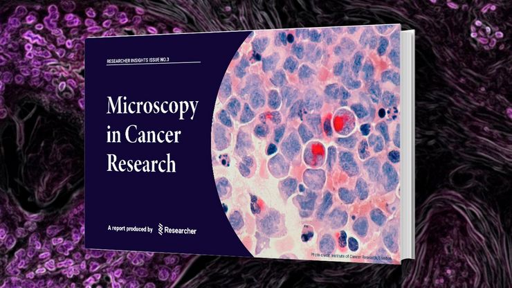
Researchers Insights: Microscopy in Cancer Research
Discover how imaging techniques are driving cancer research forward. In this issue, we present comprehensive multimodal studies using microscopy, as well as new directions in intraoperative cancer…
, Tropomyosin (cardiomyocytes, red) and GFP (primordial cardiac layer, green).")
显微镜中的荧光
荧光显微技术是一种特殊的光学显微镜技术。它利用的是荧光色素在一定波长的光激发下发光的能力。通过抗体染色或荧光蛋白标记,可以用这种荧光色素标记感兴趣的蛋白质。这样就可以确定单分子物种的分布、数量及其在细胞内的定位。此外,还可以进行共定位和相互作用研究,使用可逆结合染料(如 Ca2+ 和 fura-2)观察离子浓度,以及观察细胞的内吞和外吞过程。如今,利用荧光显微镜甚至可以对亚分辨率颗粒进行成像。

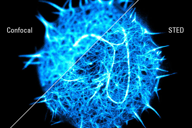
超分辨率显微镜图片库
由于光的衍射极限,传统共聚焦显微镜无法分辨约240纳米以下的结构。当需要提高分辨率以研究衍射极限尺度以下的结构和分子事件时,会使用超分辨率显微镜技术,如STED、PALM或STORM,或某些解卷积处理方法。

细胞活成像的纳米级扩展
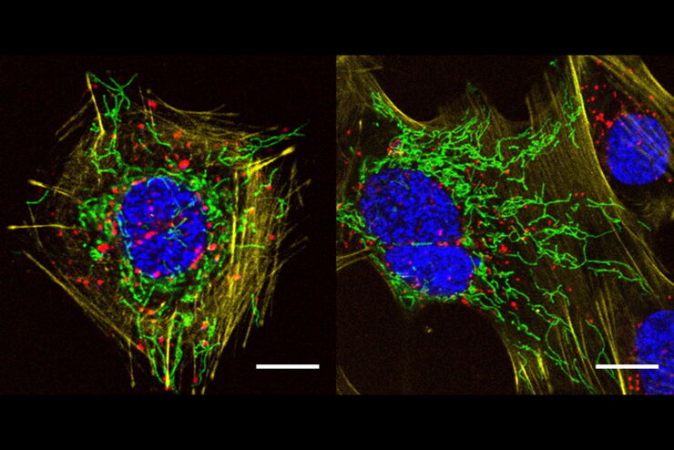
新的STED显微技术方法——TauSTED Xtend,使得在纳米级别下对活体完整样本进行扩展多色成像成为可能。通过结合空间和寿命信息,TauSTED Xtend提供了额外一层信息,允许在极低的光剂量下分辨小细节并在整体结构中解析它们。
, actin network (ATTO 647N), and nuclear pore basket (CF 680R).")
STED样品制备指南
这份指南旨在帮助用户优化受激发射损耗(STED)纳米成像的样品制备,特别是在使用徕卡微系统的STED显微镜时。它提供了单色STED成像用荧光标记的概述,并对其性能进行了评级。

采用徕卡THUNDER-DM6B观察SARS-CoV-2感染宿主细胞及其复制过程
冠状病毒2致重度急性呼吸综合征(SARS-CoV-2)
冠状病毒2致重度急性呼吸综合征(SARS-CoV-2)出现于2019年末,并快速传播全世界。由于其大面积的影响,研究人员对病毒的性质进行了深入的研究以期最终阻止大流行。一个重要的方面是病毒如何在宿主细胞中复制。Ogando及其同事的研究已经揭示了SARS-CoV-2的复制动力学、适应能力和细胞病理学。他们的工具之一是用荧光显微镜观察SARS…

采用单损耗激光的五色FLIM-STED显微镜
网络研讨会,内容涉及使用单一损耗激光和荧光寿命phasor分离技术的五色STED技术。
A Versatile Palette of Fluorescent Probes
Researchers at the Max Planck Institute for Medical Research in Heidelberg have developed a general strategy to synthesize live-cell compatible fluorogenic probes, and the result are the new MaP (Max…

结合 STED 和Lifetime的优势
在这次访谈中,Alberto Diaspro教授讨论了白光激光的优势以及STELLARIS 8 STED的TauSTED技术能力。他分享了自己在使用TauSense、荧光寿命成像和相位分析技术进行科研项目时,与共聚焦系统相关的经验。
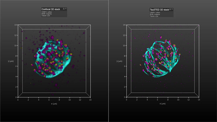
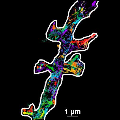
利用TauSTED在三维空间中观察有丝分裂期间的着丝粒组装
基于 TauSTED(利用寿命的受激发射损耗)技术并结合多根 STED 线(592、660 和 775 纳米),可以呈现有丝分裂纺锤体的三维组织,以及 CENP-C 和 BUB1 的分布情况,从而为着丝粒组装提供深入见解。

Regulators of Actin Cytoskeletal Regulation and Cell Migration in Human NK Cells
Dr. Mace will describe new advances in our understanding of the regulation of human NK cell actin cytoskeletal remodeling in cell migration and immune synapse formation derived from confocal and…
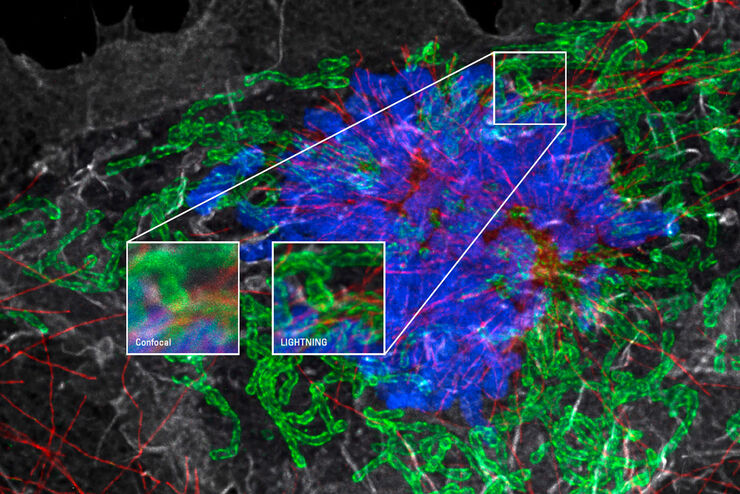
使用 LIGHTNING 可从样本中获得丰富的信息
LIGHTNING 是一个自适应的信息提取过程,可以完全自动化地呈现原本不可见的微小结构和细节。 与为整个图像使用全局参数集的传统技术不同,LIGHTNING 为每一个像素计算一个适当的参数集,尽力还原细节。
, and trafficking vesicles labeled with CF594 (cyan - Biotium).")
超高解析度
徕卡显微系统所提供的超高分辨率显微镜,通过宽场(GSD)和共聚焦(STED)技术克服了衍射极限,是您能够进一步研究亚细胞结构和动态,而这一层级的观察在之前采用普通荧光显微镜是无法实现的。
超分辨率 STED 光谱学
分子相互作用在细胞信号传导中至关重要。它们通常受到所涉及分子的流动性的影响。
通用 PAINT – 动态超分辨率显微镜
超分辨率显微技术在过去十年中彻底革新了生物学研究。这些技术让我们能够以接近蛋白质大小的分辨率观察细胞内的各个组成部分。然而,对活细胞进行成像仍然是大多数超分辨率技术面临的挑战。在这种背景下,uPAINT(纳米尺度拓扑成像通用点积累)技术受到了广泛关注。这种单分子方法通过动态成像活细胞中持续标记的任意膜生物分子,实现了超高分辨率成像,并能追踪单个分子的运动轨迹。
